< back
Branch retinal vein occlusion
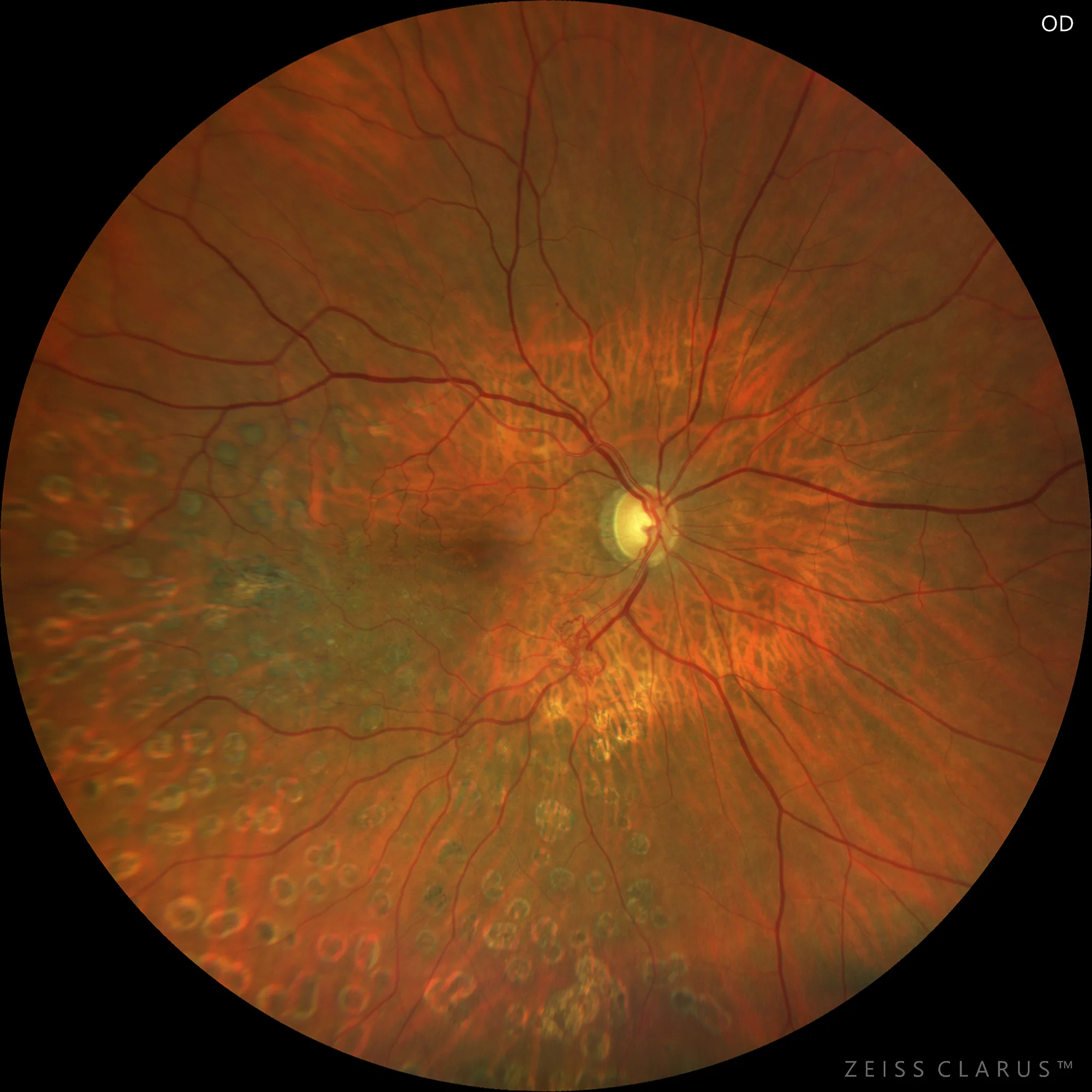
WF color retinography (Clarus 700): ORVR with sectorial panphotocoagulation and vascular alteration in the lower temporal arch
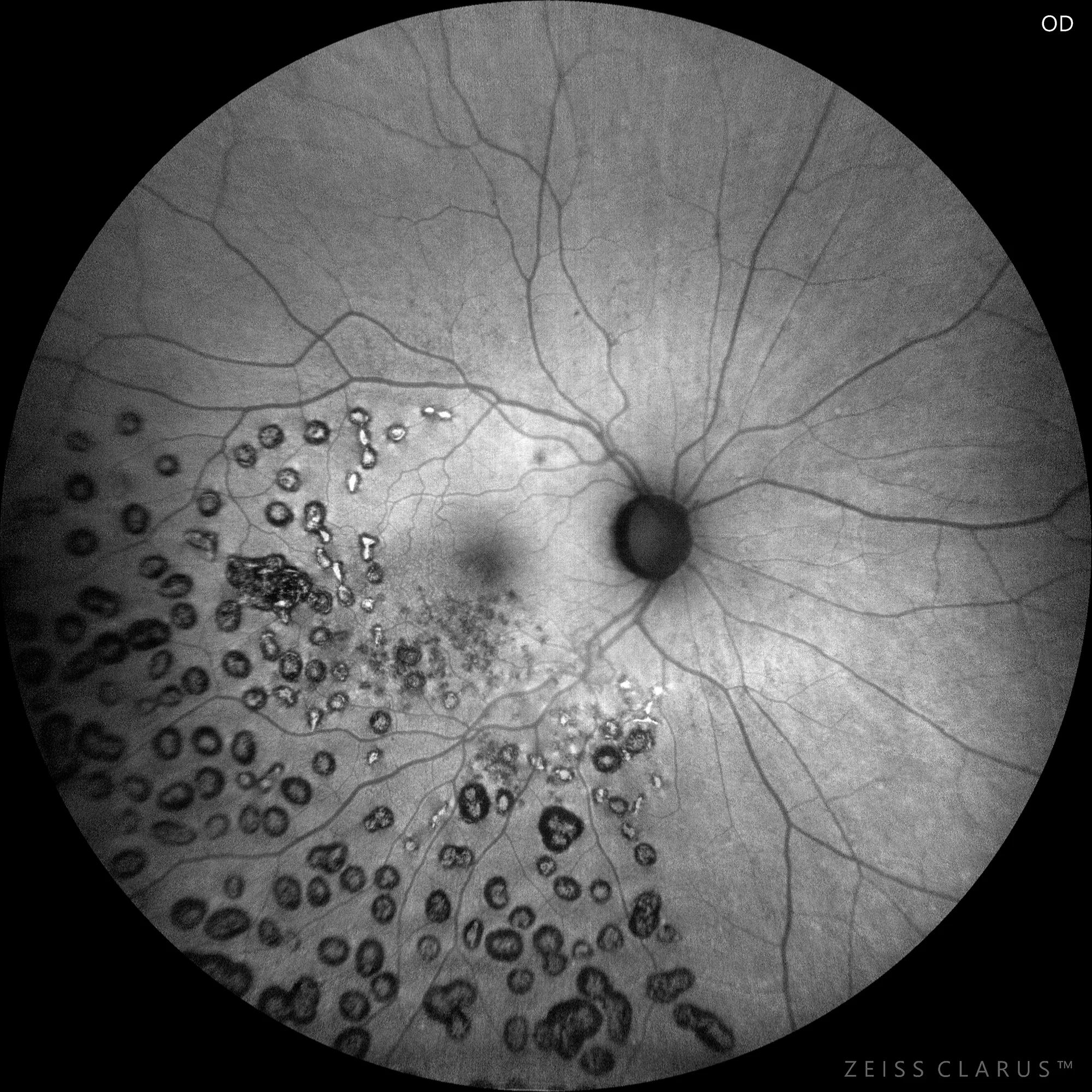
Background Autofluorescence (AF Green, Clarus 700): Allows a better appreciation of the panphotographically-agulated area
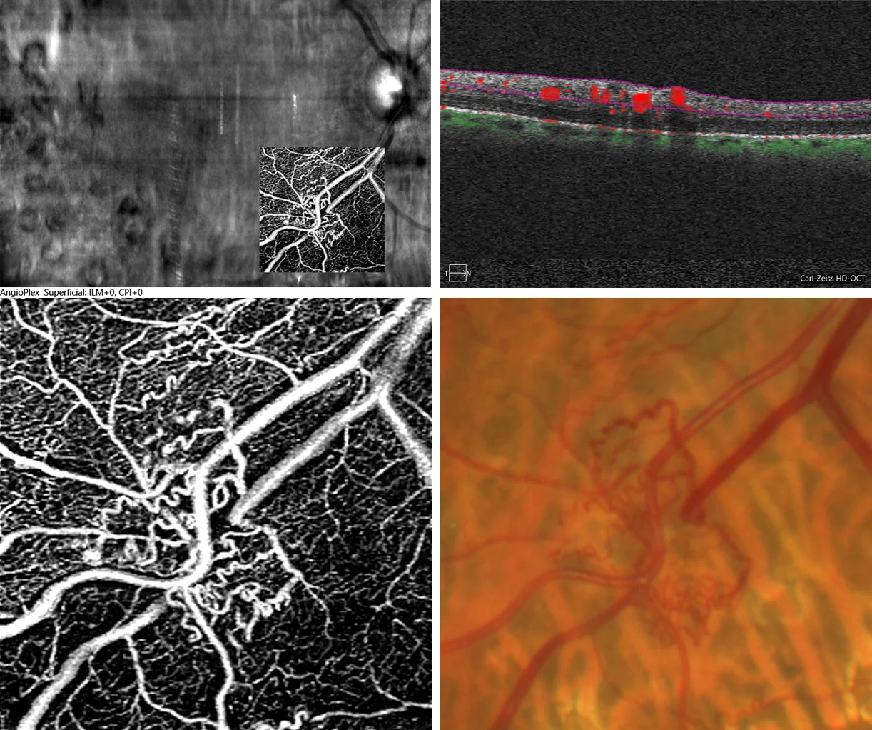
AngioOCT 3 x3 (Angioplex, Zeiss): Allows confirmation of the intraretinal location of vascular shunts. Enlarged detail of retinography.
Description
Branch retinal vein occlusion (BRVO) is the second most common vascular disorder after diabetic retinopathy. The main complications of BRVO are the presence of macular edema and neovascularization. It is important to differentiate the presence of neovessels from vascular shunts, which can be difficult to differentiate during fundus examination.