Cavernous Hemangioma
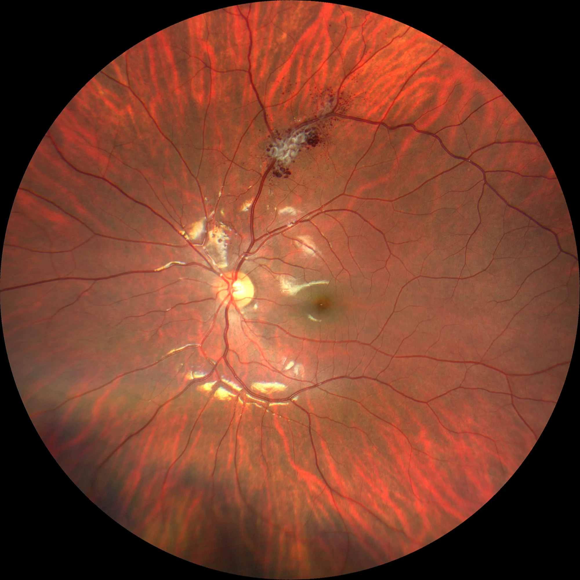
Color retinography LE: Cavernous hemangioma in the upper temporal vascular arcade. The grape-shaped saccular angiomatous lesions can be observed.
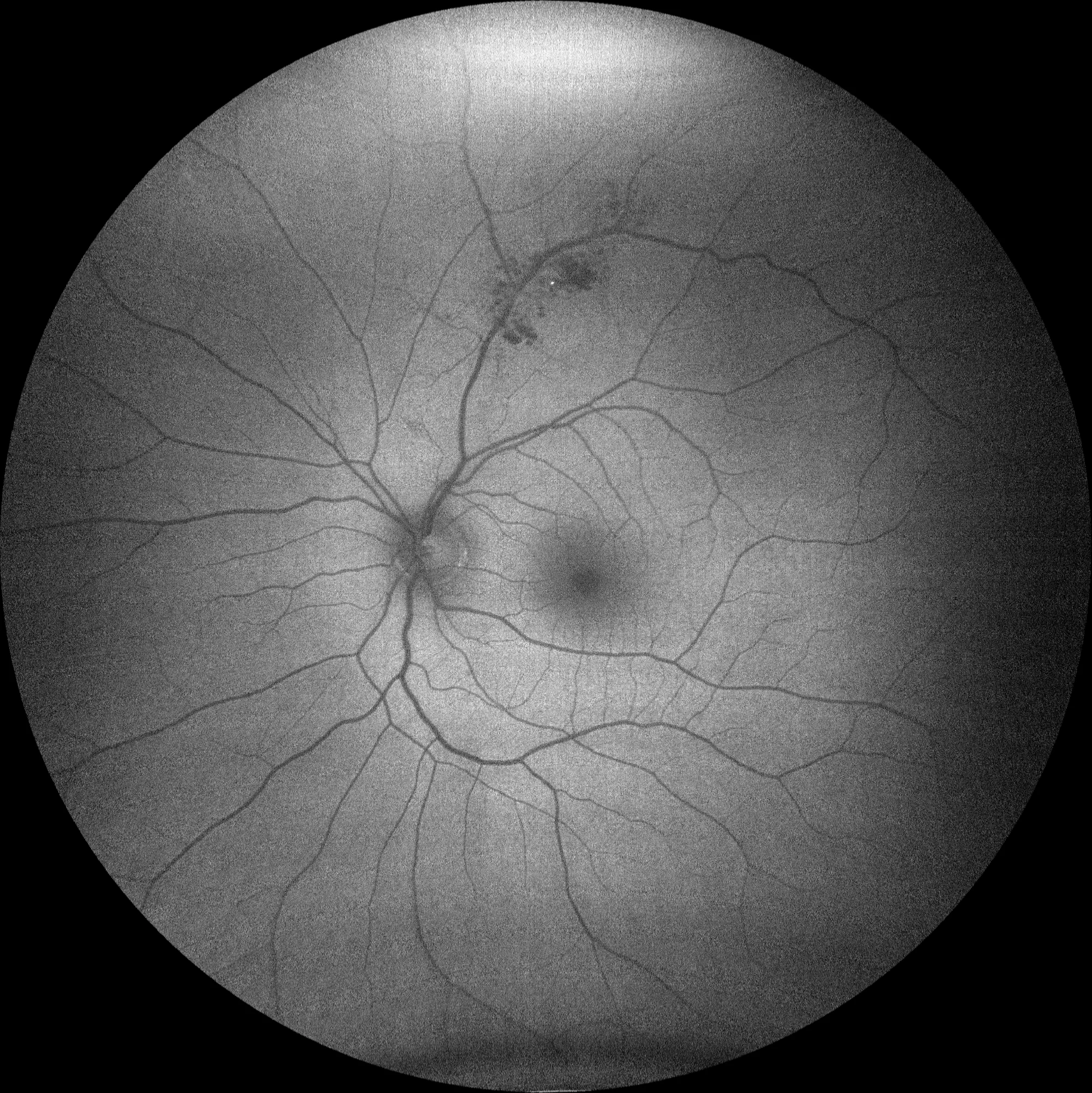
OD: Autofluorescence shows hypoautofluorescent lesions corresponding to saccular vascular formations that are part of the hemangioma.
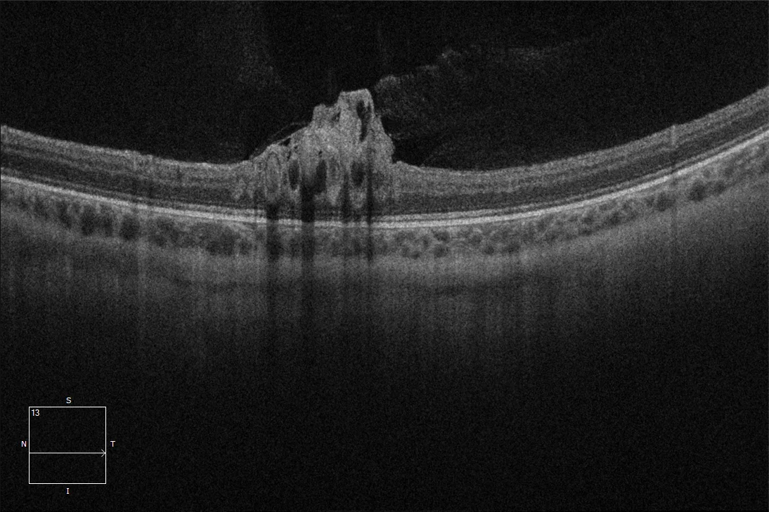
OCT on the OD lesion: In the OCT we can observe a hyperreflective lesion located in the inner retina that contains hyporeflective areas inside that correspond to vascular aneurysms.
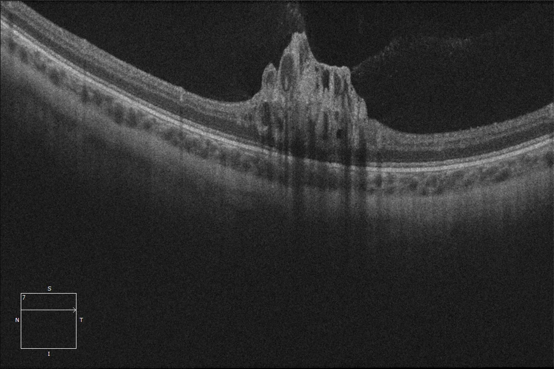
OCT on the OD lesion: In the OCT we can observe a hyperreflective lesion located in the inner retina that contains hyporeflective areas inside that correspond to vascular aneurysms.
Description
Cavernous hemangioma is a benign vascular tumor of the retina or optic nerve head. It is usually sporadic, although it may have a familial pattern (autosomal dominant) that may be associated with hemangiomas of the central nervous system and skin. The lesions are characterized by the formation of saccular angiomatous lesions that have the appearance of a bunch of grapes. They are usually small and asymptomatic so they do not require treatment, although occasionally hemovitreous or fibroglial formations that pull on the retina may occur.