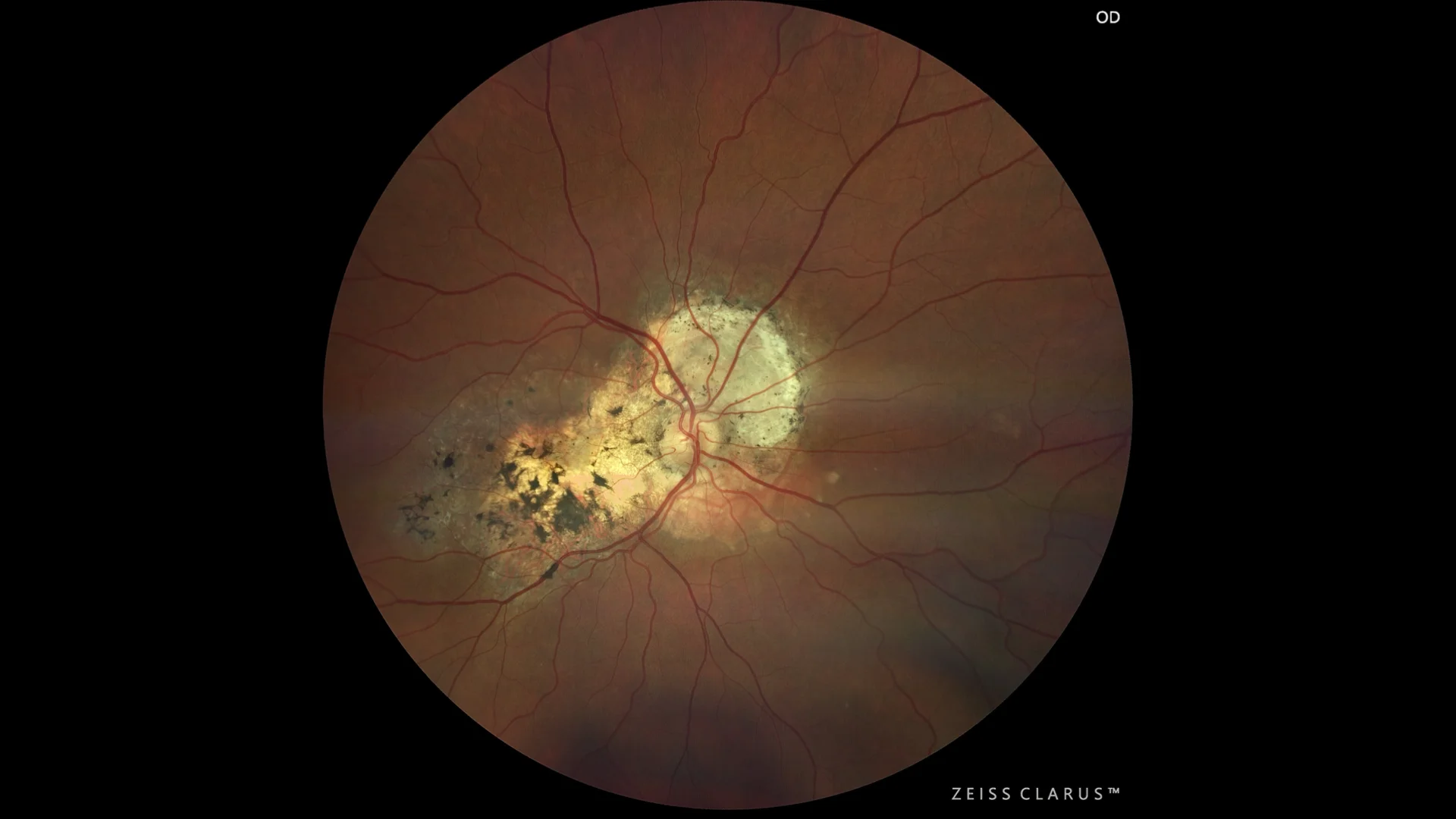
Choroidal osteoma
Irregular lifting of the RPE, with medium hyperreflectivity, with a laminar pattern.
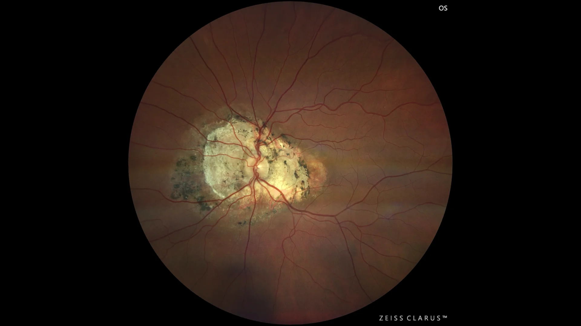
Yellowish-whitish lesion, the margins show some pigmentation and atrophy. It extends from the peripapillary area towards the macula.
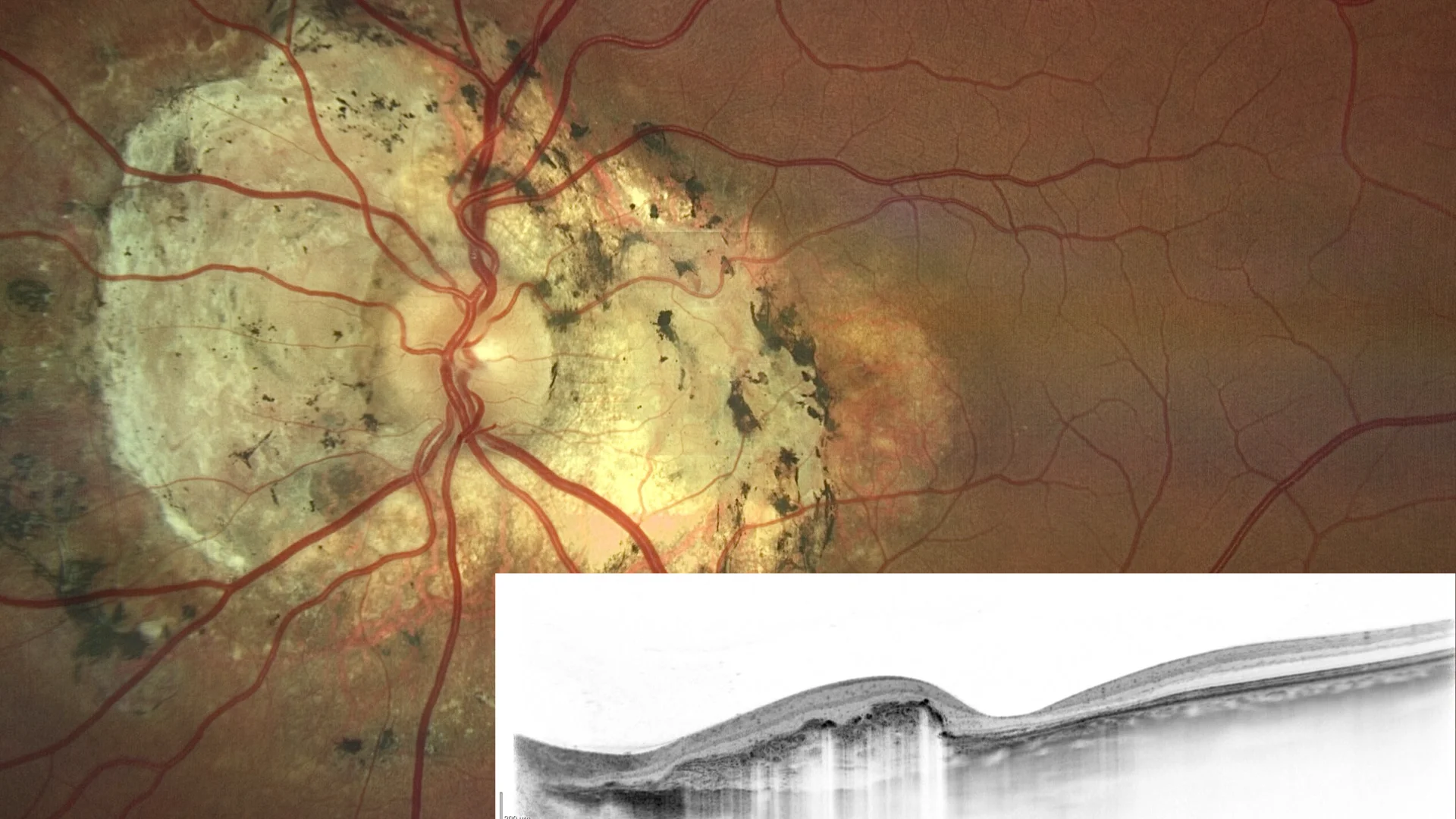
OCTsd: with irregular elevation of the RPE, showing a laminar pattern.
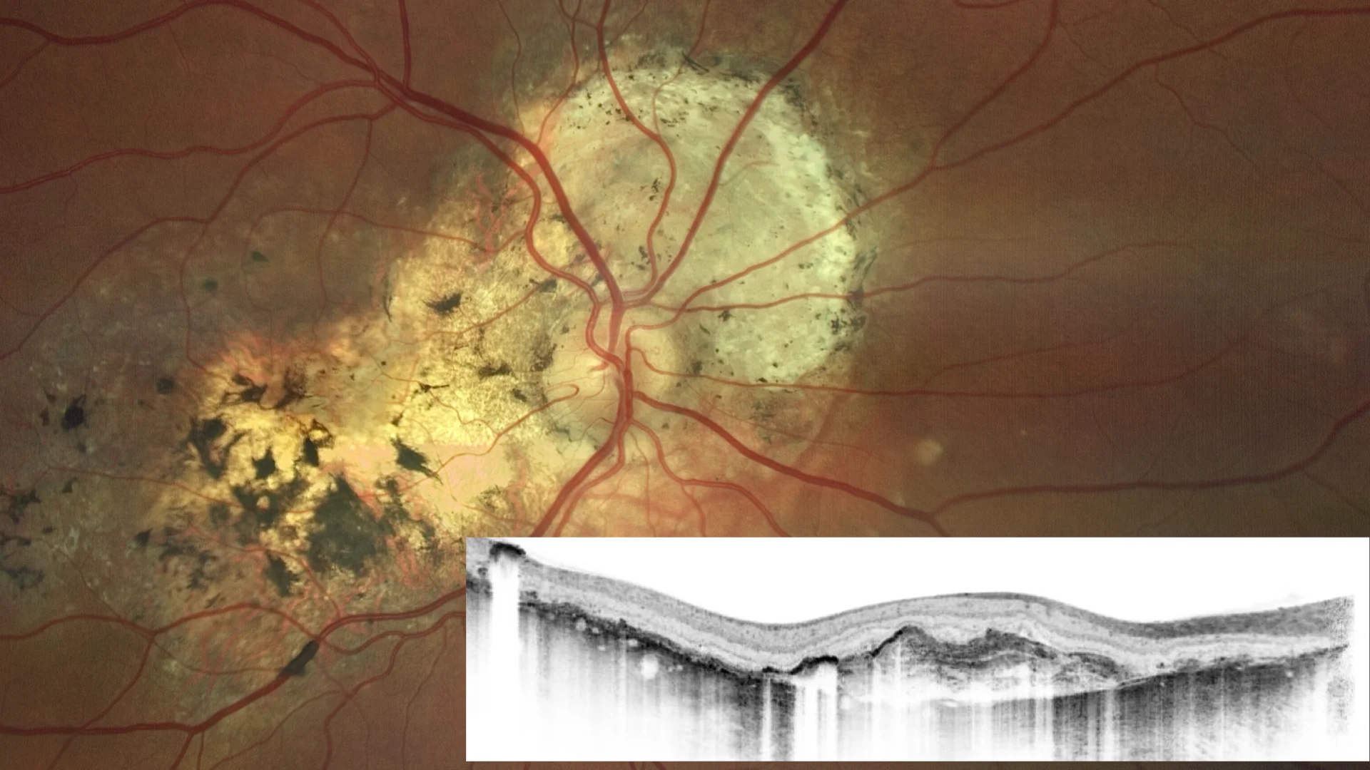
OCTsd: with irregular elevation of the RPE, showing a laminar pattern, and respect for the subfoveal area.
Description
Choroidal osteoma is a rare, benign ocular tumor, primarily composed of mature bone, that forms in the choroid.
It is most often diagnosed in young women and adolescents, although its exact cause is unknown. This tumor can be solitary or multiple, and tends to be located near the optic nerve or in the macular region. Often, choroidal osteomas are asymptomatic and are discovered incidentally during a conventional examination, but they usually end up affecting vision. An ultrasound, X-ray, or CT scan can be performed to confirm the calcification of the osteoma, although OCT is usually very useful. Treatment is controversial, and unless choroidal neovascularization develops, antiVEGF drugs are not useful.