Choroidal osteoma
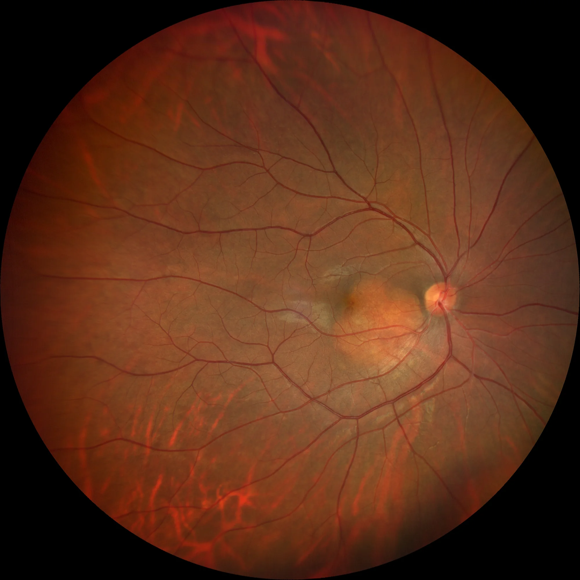
Color retinography (Clarus500, Zeiss): (A) Color retinography OD: Orange choroidal lesion located in the papillo-macular bundle affecting the macula, which corresponds to a choroidal osteoma.
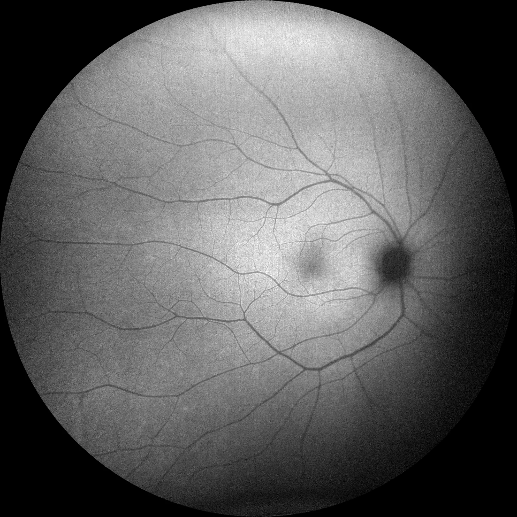
Autofluorescence (Clarus500, Zeiss): (B) OD: Autofluorescence shows subtle hyperautofluorescence in the tumor area. Since the lesion has not yet decalcified and is not causing alterations in the retinal pigment epithelium (RPE) or the overlying retina, no significant alterations are seen in the autofluorescence. Choroidal osteomas frequently cause alterations in the RPE that result in hyper-hypoautofluorescence in the autofluorescence.
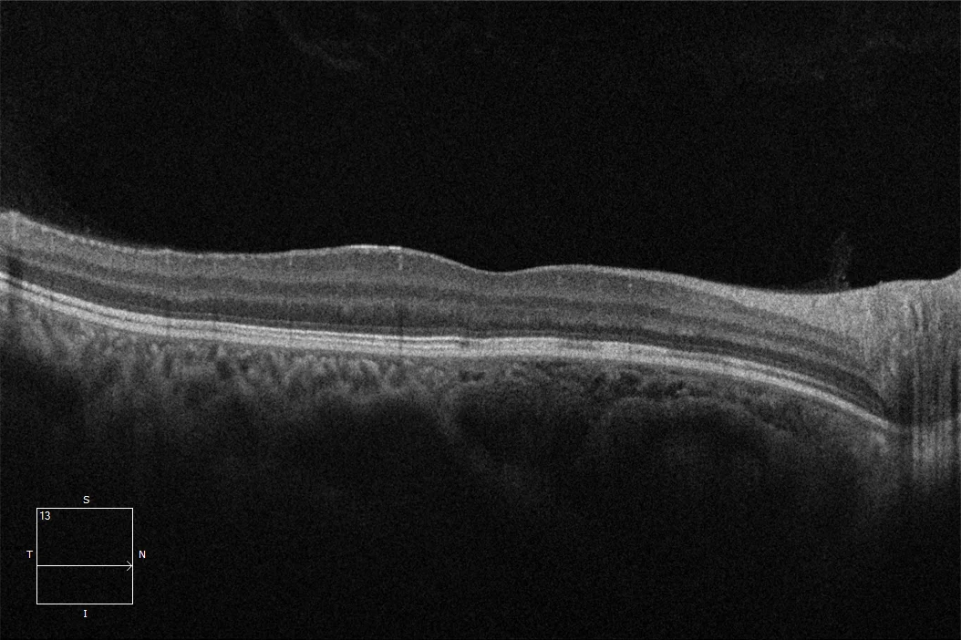
Macular OCT OD: OCT shows a space-occupying choroidal lesion in the papillo-macular bundle that reaches the fovea and has a different structure than the choroid.
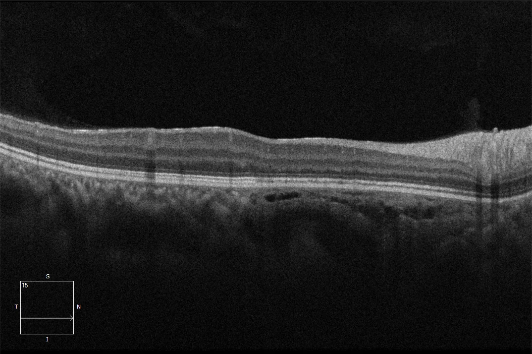
OCT over the OD lesion: Space-occupying choroidal lesion with multiple intralesional layers, spongiform appearance and intralesional vessels.
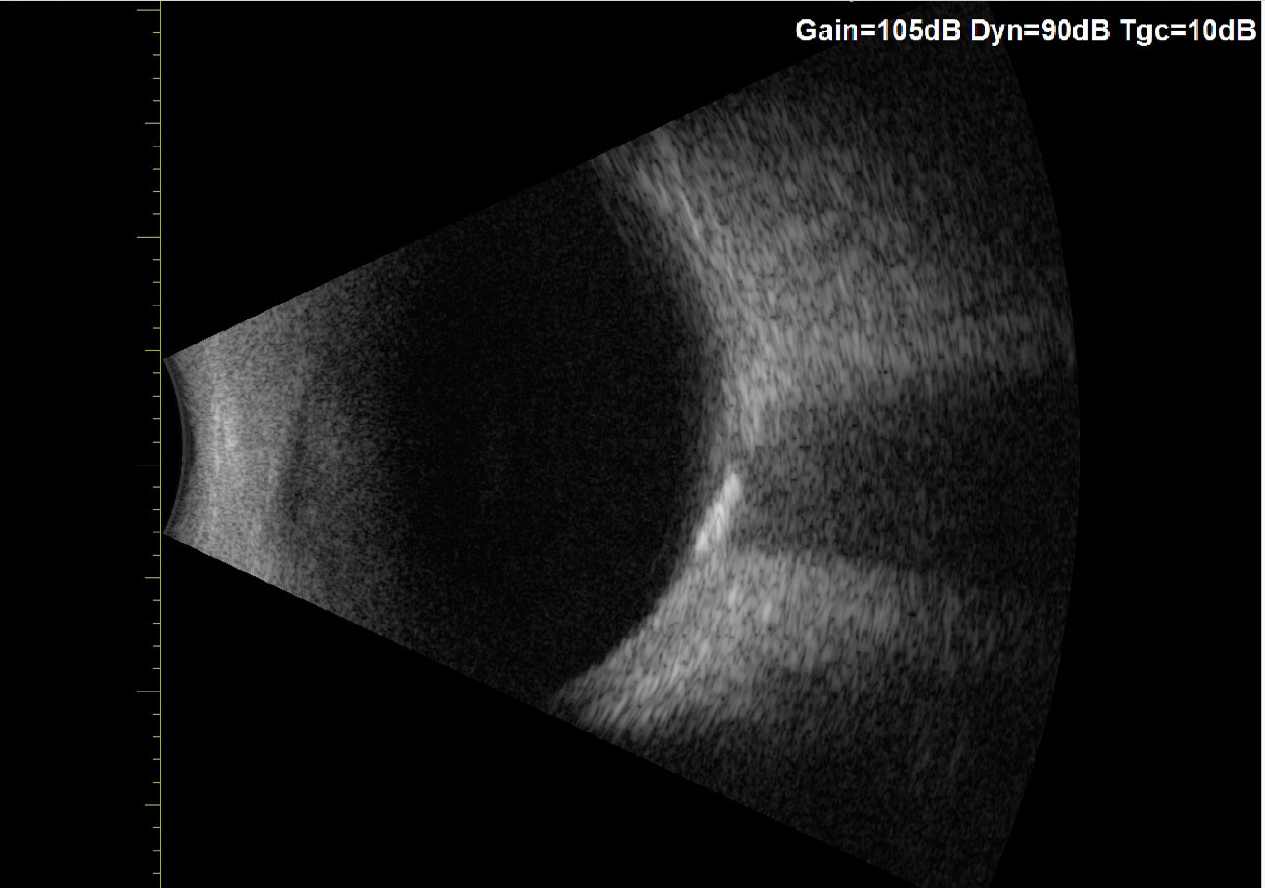
Ultrasound of the lesion: A hyperechoic choroidal lesion with posterior shadow is evident, due to the bone structure of the tumor.
Description
Choroidal osteoma is a benign choroidal tumor composed of mature bone tissue that is usually located in the juxtapapillary region. This tumor has the ability to form authentic bone tissue with trabeculae and vascularized bone marrow in the choroid. It typically appears in young women in the second-third decade of life, and usually occurs sporadically and unilaterally (75%). It usually presents as an oval, orange, well-defined lesion, which may be slightly raised (0.5-3 mm). The basal diameter of these tumors may increase over the months or years, extending towards the macula, so their visual prognosis is variable and unpredictable since it will depend on the macular involvement. Extrafoveal osteomas usually maintain good visual acuity, while those that affect the fovea may suffer severe vision loss generally caused by atrophy or alteration of the RPE and photoreceptors, the presence of subretinal fluid or the appearance of neovascular membranes. The management of these tumors is based on preventing tumor growth towards the fovea. Photodynamic therapy has proven to be effective in this regard by inducing a process of decalcification and partial regression of the tumor that stabilises it, so it may be indicated in extramacular tumors growing towards the fovea.