Choroidal tuberculomas
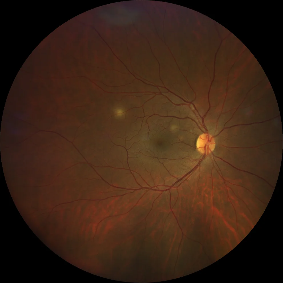
A. Color retinography (Clarus 500, Carl Zeiss Meditec ASG, Jena, Germany) showing multiple deep, yellowish lesions with diffuse edges in the posterior pole.
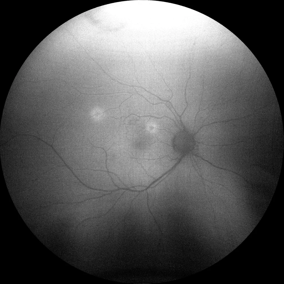
B. Autofluorescence image (Clarus 500, Carl Zeiss Meditec ASG, Jena, Germany) showing hypoautofluorescent lesions with hyperautofluorescent halo.
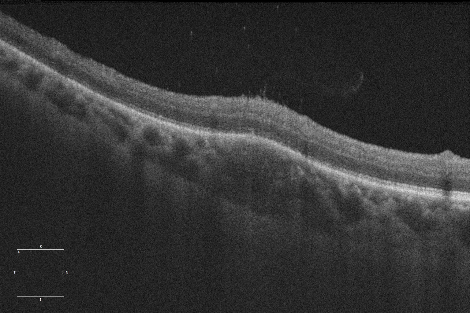
C. Macular optical coherence tomography (Cirrus 5000, Carl Zeiss Meditec ASG, Jena, Germany) showing a hyperreflective lesion in the choriocapillaris with bulging of the pigmented epithelium and overlying neurosensory retina, as well as hyperreflective spots in the vitreous overlying the lesion.
Description
Ocular tuberculosis is a form of extrapulmonary involvement of tuberculous infection. It can involve all ocular structures, but most frequently affects the uvea, and the pathophysiology is directly infectious or of autoimmune origin. One of its classic presentations is in the form of choroidal tubercles and tuberculomas, true caseating granulomas that appear as yellow-grayish lesions preferably in the posterior pole and middle periphery. In autofluorescence, lesions with a central hypoautofluorescent nucleus can be observed, which corresponds to the area of necrosis surrounded by a hyperautofluorescent inflammatory halo with poorly defined edges. In optical coherence tomography, the presence of hyporeflective lesions in the choriocapillaris with overlying subretinal fluid in the outer layers of the retina is characteristic.