Chronic central serous chorioretinopathy
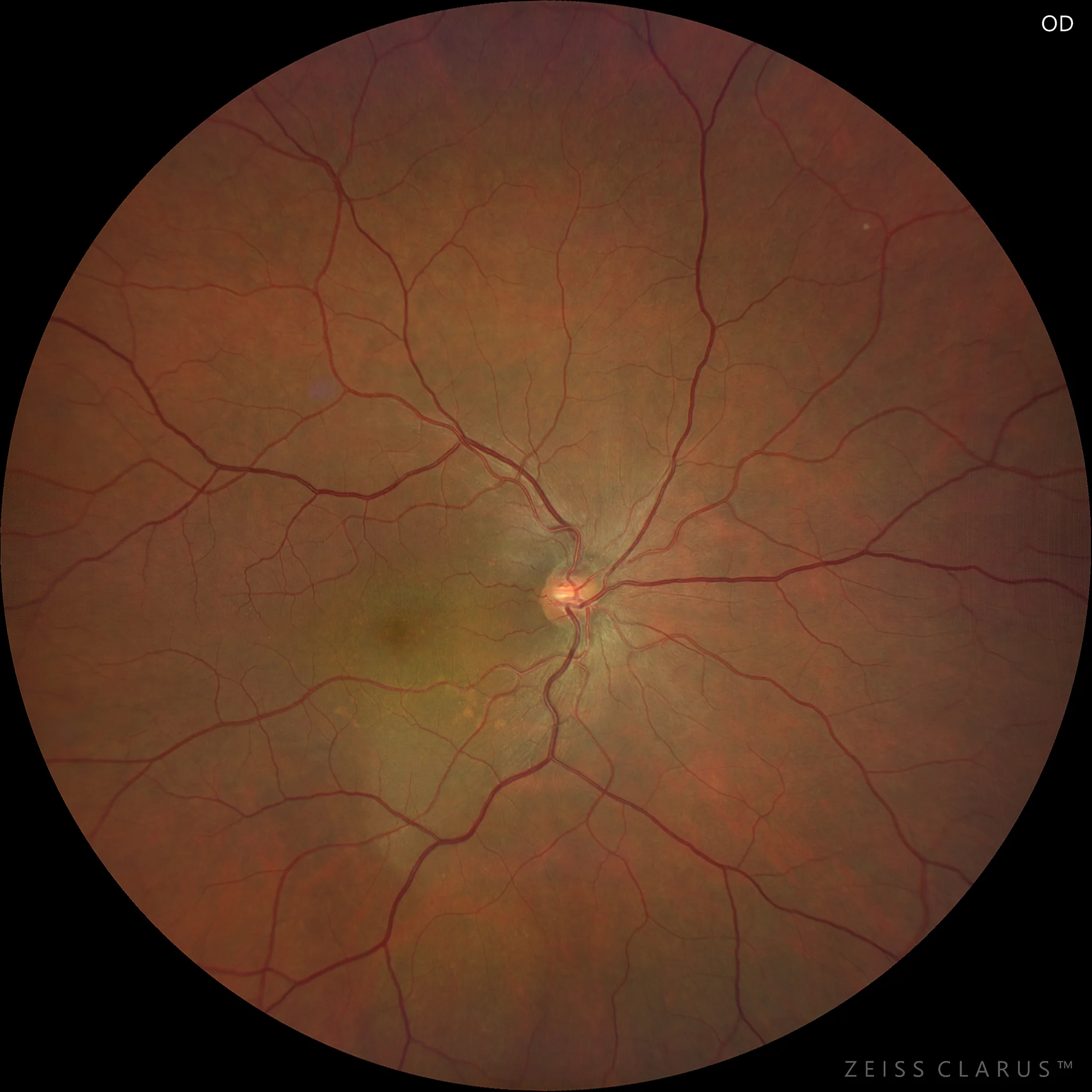
Color retinography showing a yellowish-green stain in the lower macular region. Accumulations of orange pigment are observed.
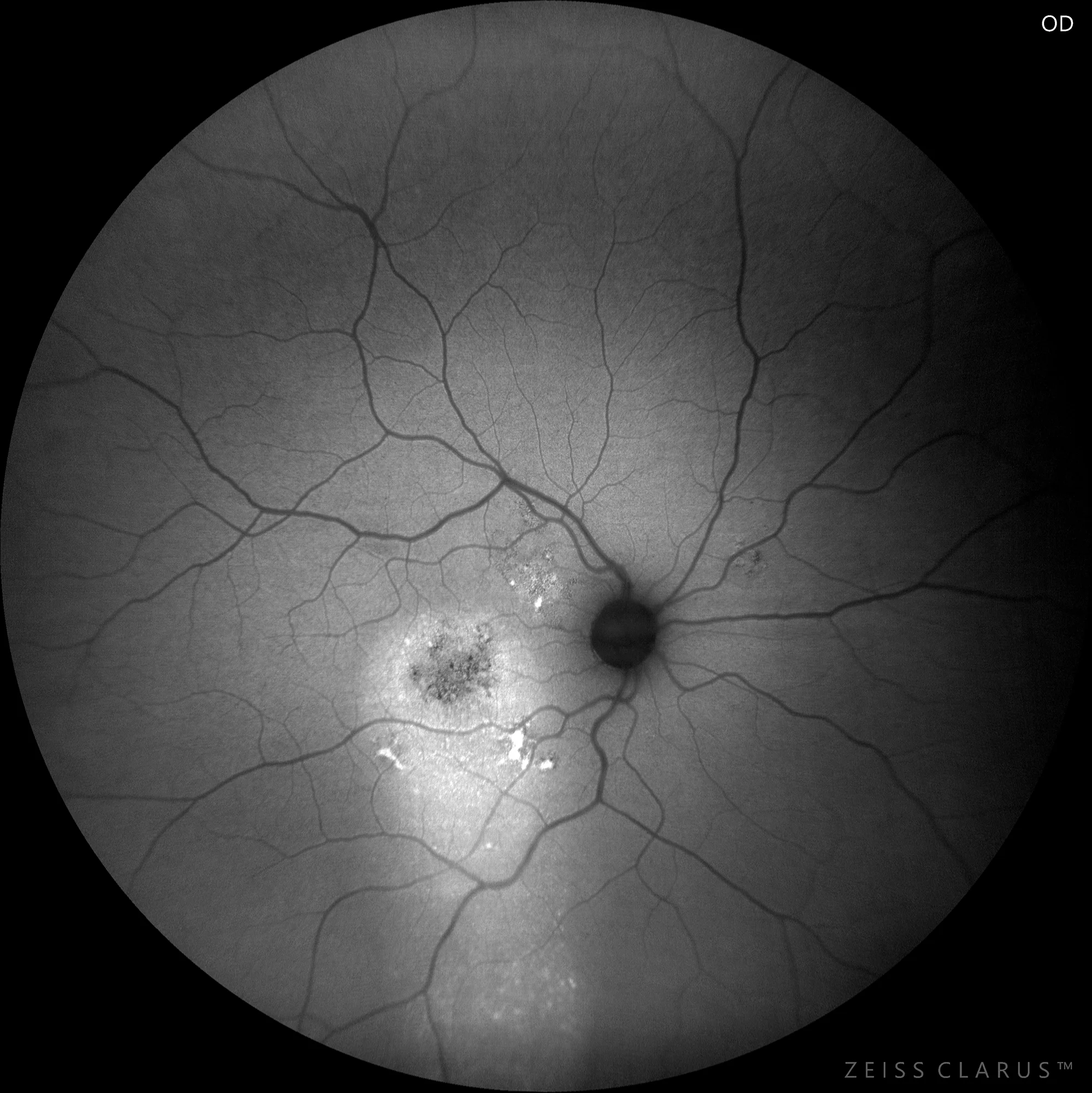
Green autofluorescence: a main lesion is observed in the macula, with a gradual descent of the macula in a stream, and with hyperautofluorescence of the orange pigment deposits. A hypoautofluorescent mottling is observed in the macula, indicating diffuse atrophy of the RPE. Another much smaller focus is observed superonasally to the macula.
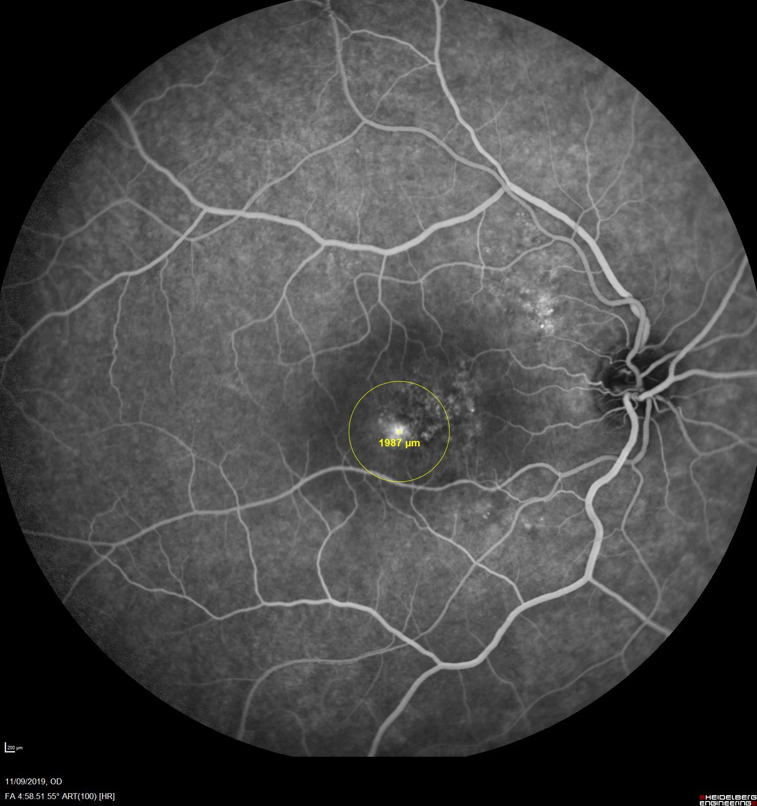
Fluorescein angiography shows hyper/hypofluorescent mottling in the macular region, with a clear focus of staining and diffusion. The image reflects the treatment spot for PDT.
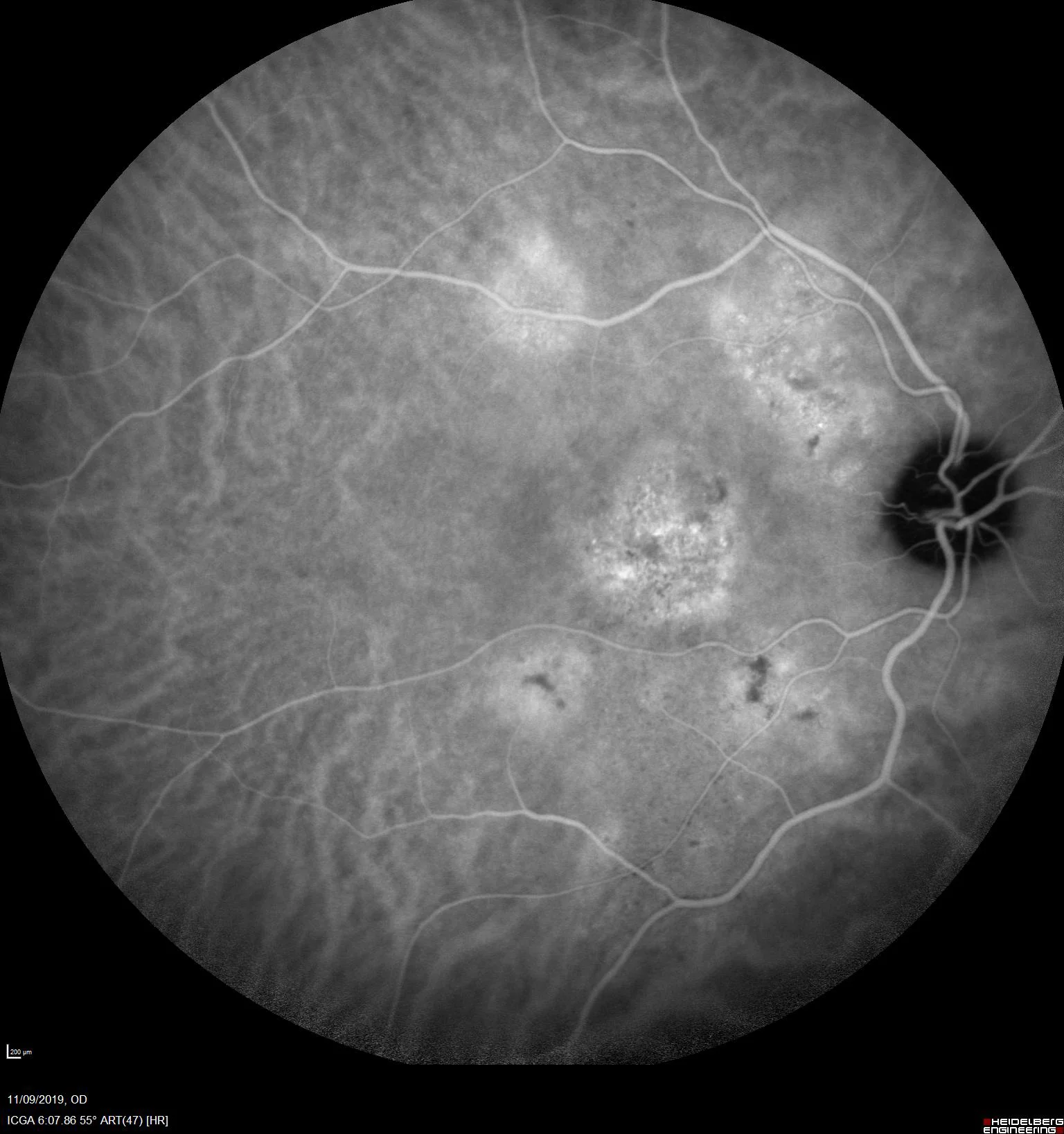
Indocyanine green shows multiple hypercyanescent plaques at medium times. Lipofuchsin accumulations appear hypocyanescent due to the screen effect.

OCT shows a neurosensory detachment, along with a flat bilobed PED, and hyporeflective content. In turn, the presence of large-calibre choroidal vessels in an anterior position can be noted in the choroid, which may apparently compress the choriocapillaris.
Description
Chronic central serous chorioretinopathy (CSCR) is a condition characterized by persistent accumulation of subretinal fluid in the macular region resulting from dysfunction of the retinal pigment epithelium (RPE) and the outer blood-retinal barrier. On optical coherence tomography (OCT), CSCR is manifested by serous detachment of the less dome-shaped neuroepithelium and occasionally RPE detachments (RPE). Fluorescein angiography (FA) typically shows leaky spots, along with more diffuse hyperfluorescent plaques. On indocyanine green angiography (ICG), placoid hyperfluorescence is seen, indicating patches of choroidal hyperpermeability.