Coats disease
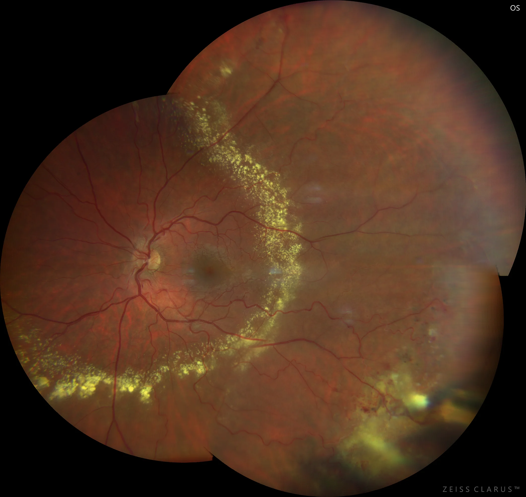
Colored retinography showing the superficial retinal plexus of normal caliber, which would differentiate it from a retinal capillary hemangioma. It shows an accumulation of lipid exudation in the form of a circinate threatening the posterior pole. In the infero-temporal quadrant we see that the exudative process is accentuated, and in turn a possible DVT is intuited.
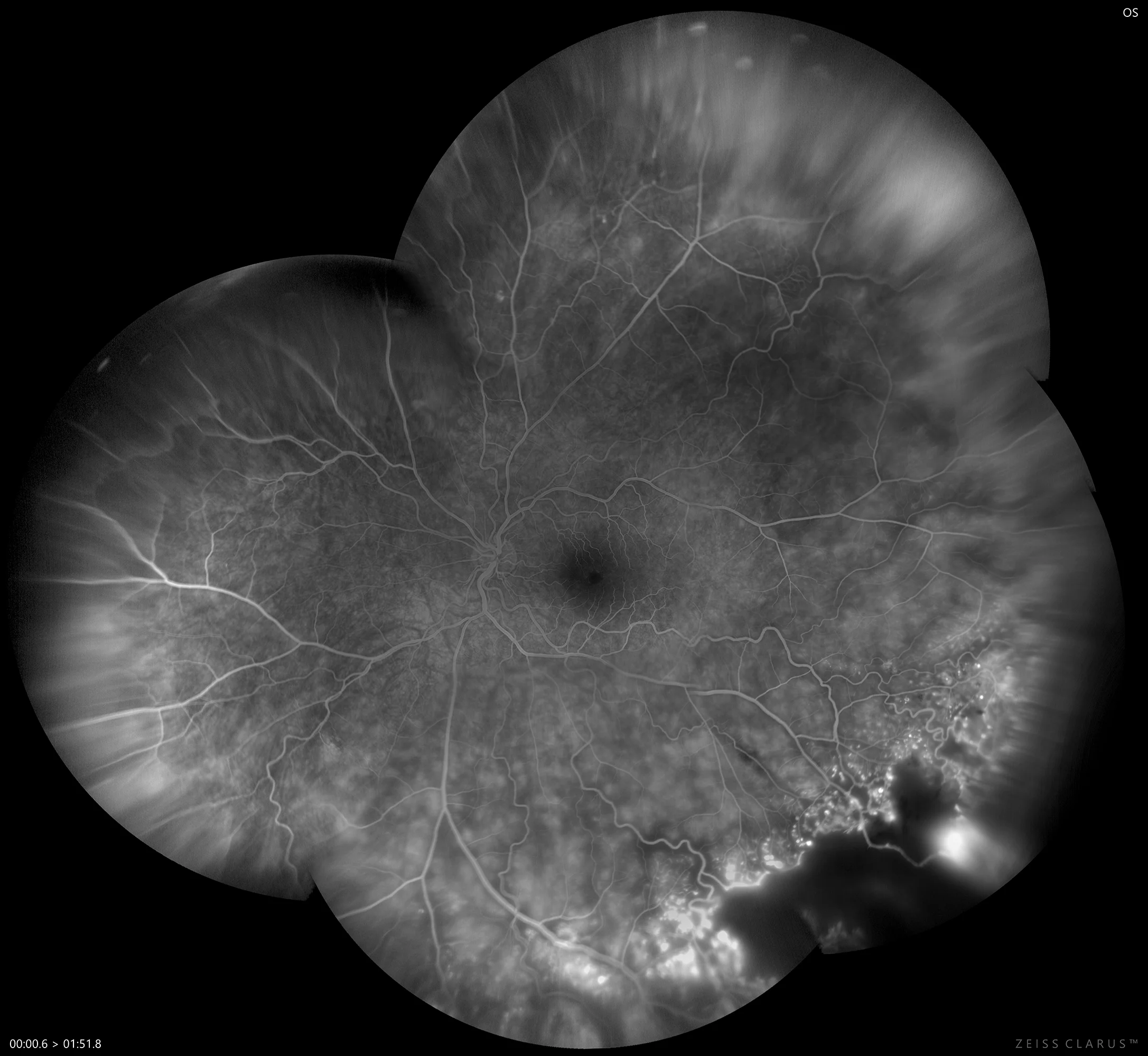
AGF: reveals multiple garland-like aneurysmal dilatations in the inferotemporal quadrant. Marked hyperfluorescence of a medium-sized nodular lesion in this quadrant is also observed, which appears to correspond to the DVT. The vascular alterations spread to the upper quadrant, although with less intensity.
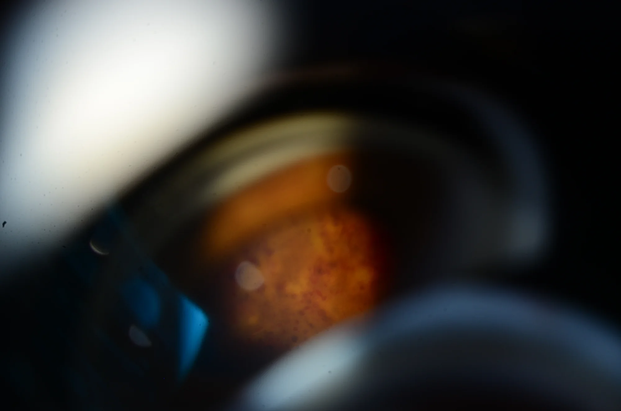
Direct visualization with the three-mirror lens, in which aneurysmal dilatations are seen in detail.
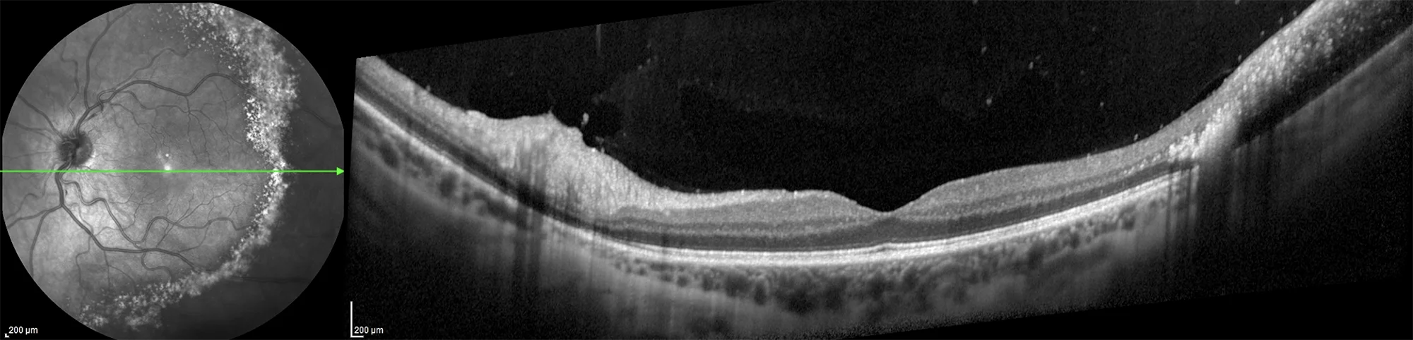
In an OCT section we can see that a pathological deposit of lipid exudation has occurred, which flows towards the outer retina. There is no macular edema or significant alteration in the vitreous-retinal interface.
Description
Coats disease is a non-inherited condition characterized by the presence of unilateral retinal telangiectasias, exudation, and exudative retinal detachment, mainly in young patients. The disease is more common in males and usually occurs unilaterally, with an average age of onset between 2 and 8 years. Arteriolar telangiectasias cause subretinal and intraretinal exudation of fluid and lipids, which may eventually lead to exudative retinal detachment and vision loss.