Collateral branches in macular venous branch occlusion
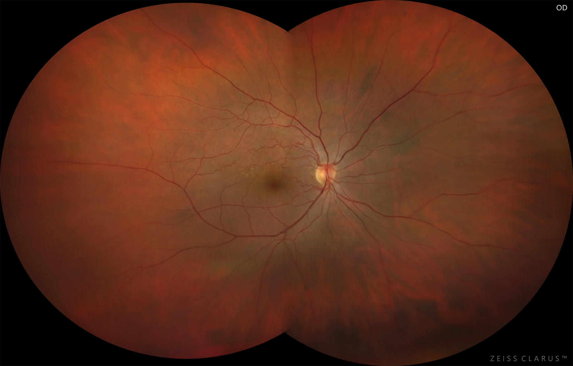
Figure 1. Color retinography of the right eye, showing collateral circulation passing through the median raphe at the level of the papillomacular bundle.
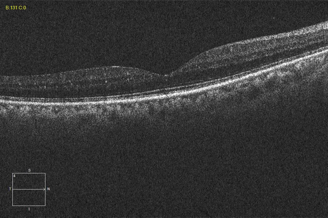
Figure 2. OCT of the right eye. Integrity of the outer retina, without macular edema.
Description
Retinal vein occlusion (RVO) is the second cause of vision loss due to retinal vascular pathology, after diabetic retinopathy. Branch vein obstruction (BRVO) is more common than central retinal vein obstruction (CRVO). Macular branch occlusion (MBO) is limited to a small branch that drains a specific sector of the macular area, with cystoid macular edema being the main cause of vision loss in these cases. The visual prognosis will depend on the location where the occlusion has occurred, the extent of ischemia, and the efficiency of collateral circulation development.