< back
Combined hamartoma of the retina and retinal pigment epithelium
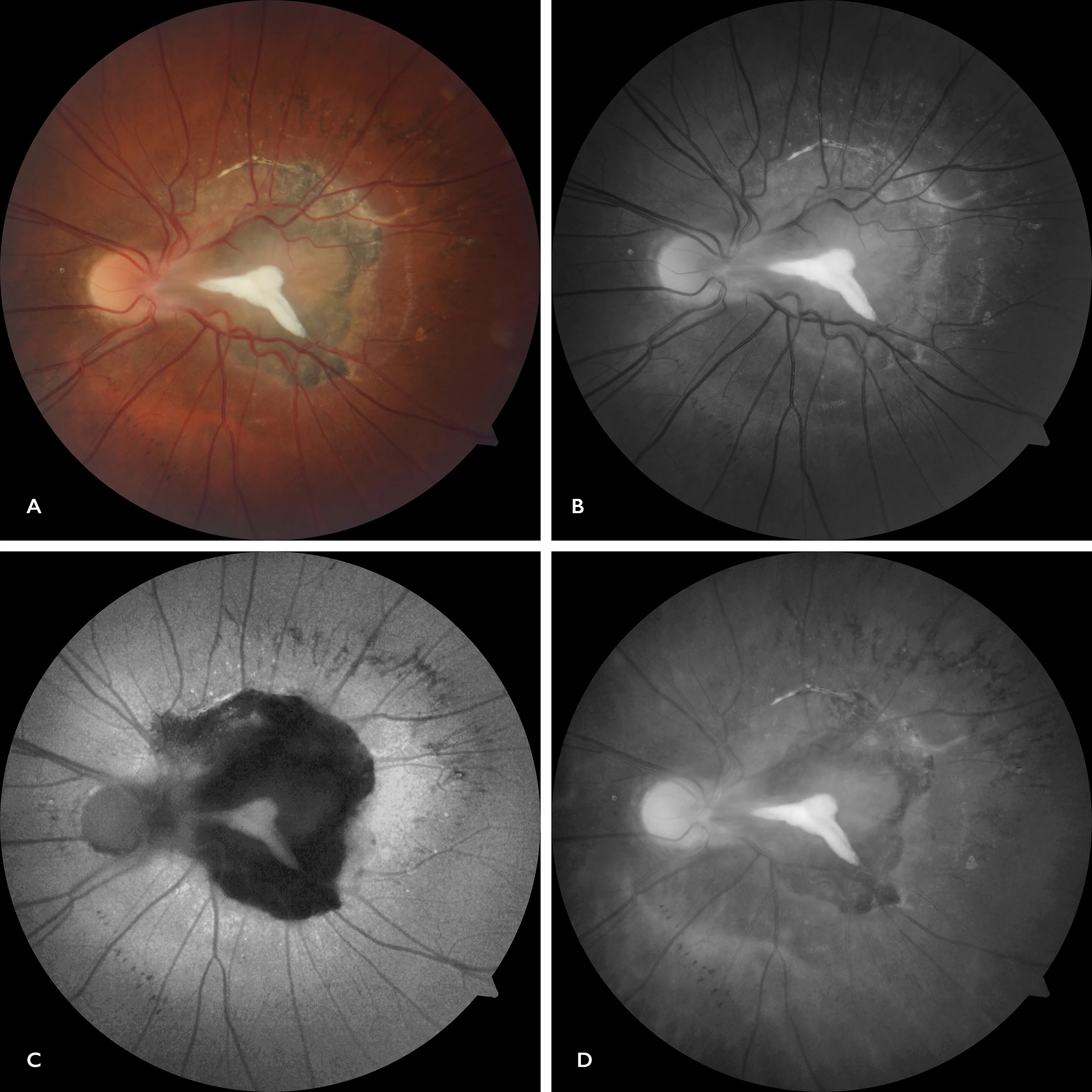
Retinography (Visucam 500, Zeiss) A: Color, B: Green filter. C: Fundus autofluorescence. D: Red filter
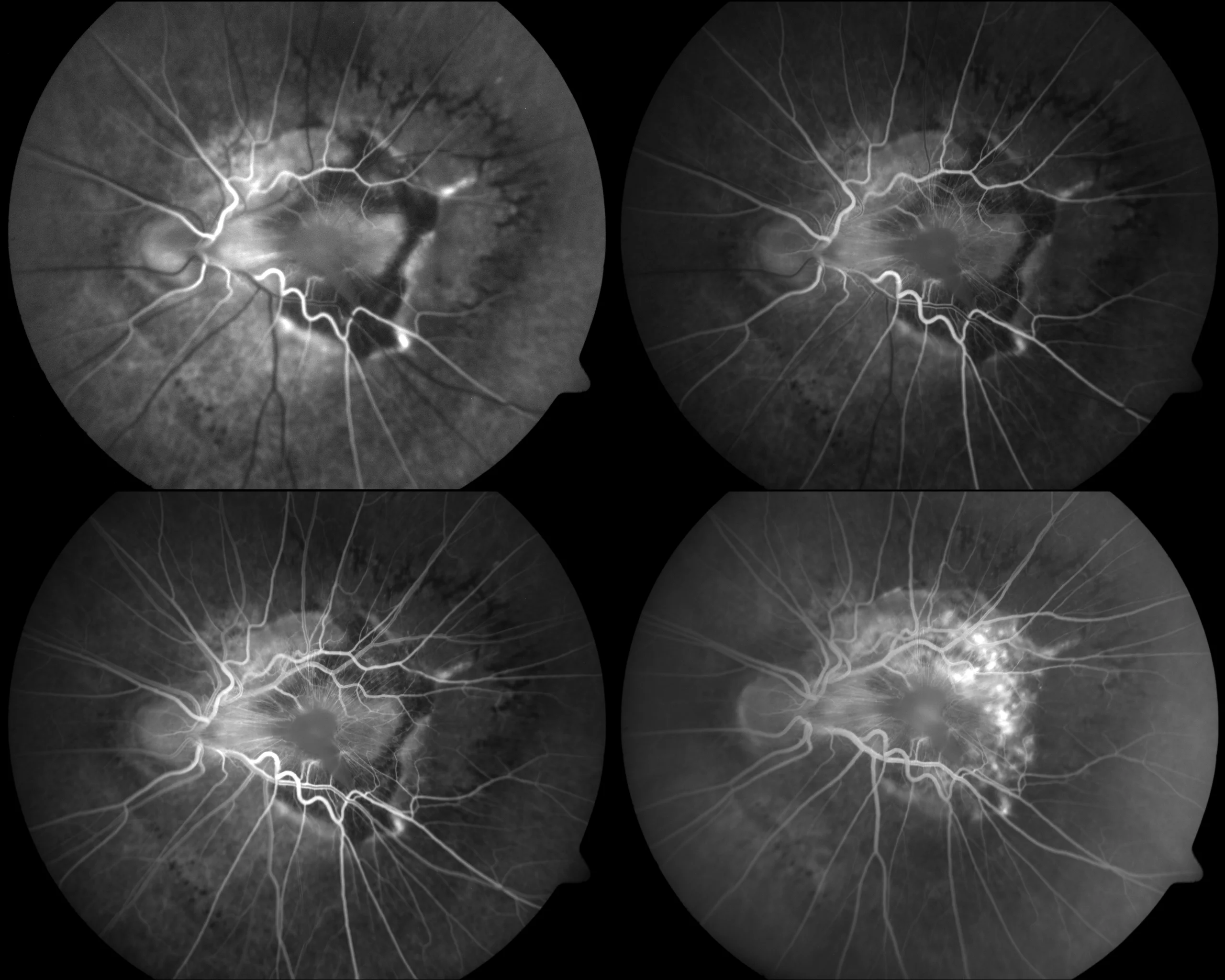
Fluorescein angiography (FF450 IR plus, Zeiss): Screen effect due to lesion pigmentation in early stages, with marked vascular tortuosity and oozing in late stages.
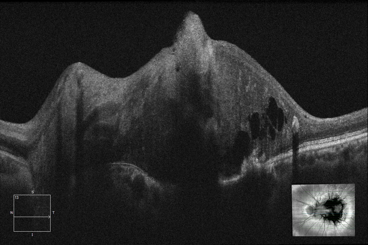
Optical coherence tomography (Cirrus-HD 5000, Zeiss): Marked thickening of the macular area with total destructuring of layers, cystic spaces and posterior shadow.
Description
Combined hamartoma of the retina and retinal pigment epithelium (HCREPR) is a benign tumor that can cause significant visual loss. HCREPR is usually a solitary, unilateral lesion located juxtapapillary or in the posterior pole. Its typical appearance is raised, vascularized, and pigmented glial tissue.
Comments
Tests:- Retinography (Visucam 500, Zeiss): Color: pigmented lesion with central whitish area. Green filter: Highlights vascular tortuosity. AF: Highlights hypoautofluorescence that delimits the lesion. Red filter: Highlights RPE alteration.
- Fluorescein angiography (FF450 IR plus, Zeiss): Screen effect due to lesion pigmentation in early stages, with marked vascular tortuosity and oozing in late stages.
- Optical coherence tomography (Cirrus-HD 5000, Zeiss): Marked thickening of the macular area with total destructuring of layers, cystic spaces and posterior shadow.