Diabetic macular edema
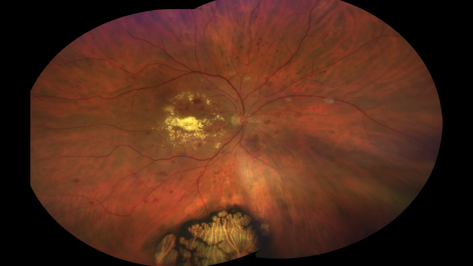
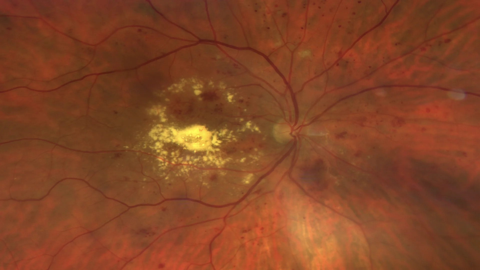
Color retinography in which hemorrhages, microaneurysms, cotton-wool exudates, atrophic changes, lipid exudation in the posterior pole and hypertrophy of the pigmented epithelium in the lower region of the retina are very evident. ๏ Color retinography magnified in the posterior pole to visualize in greater detail the exudative changes and vascular anomalies.
Optical coherence tomography in which intraretinal fluid cavities can be distinguished, as a cystoid macular edema, both in the inner and outer nuclear layers. A rounded juxtafoveal microaneurysm with hyperreflective walls can also be seen in the inner nuclear layer. In the lower image we see the same patient after two years of intravitreal treatment with anti-VEGF.
Description
Diabetic macular edema (DME) is evident on optical coherence tomography as macular thickening with hyporeflectivity due to intraretinal fluid accumulation. Lipid or circinated exudates appear as circular hyperreflective lesions, indicating microaneurysms and significant capillary leakage. These findings are essential for ophthalmologic evaluation and guide therapeutic management, which may include laser therapy, intravitreal injections of antiangiogenic agents (anti-VEGF), or corticosteroids, depending on each case.