Diffuse Choroidal Atrophy
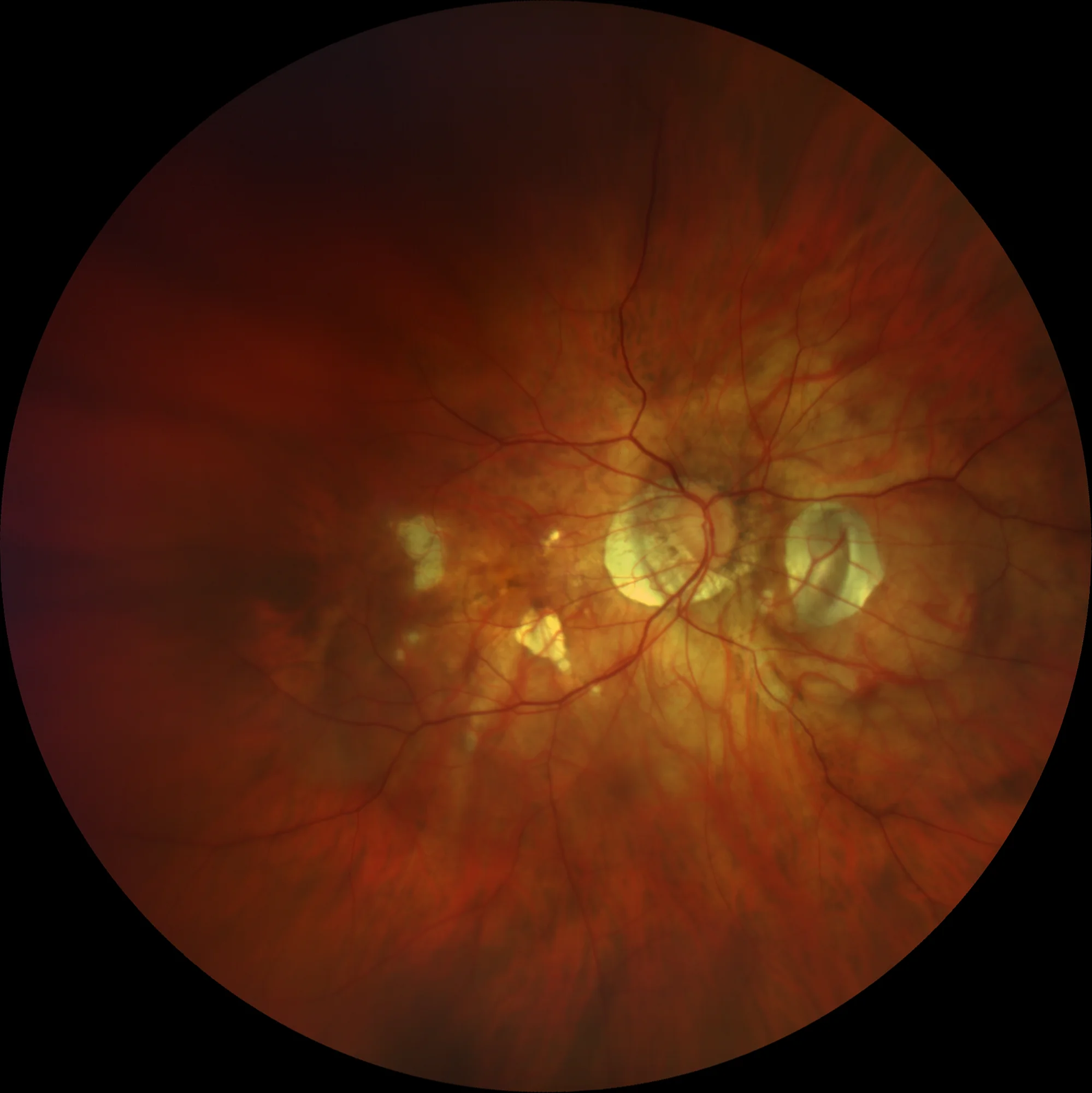
RE
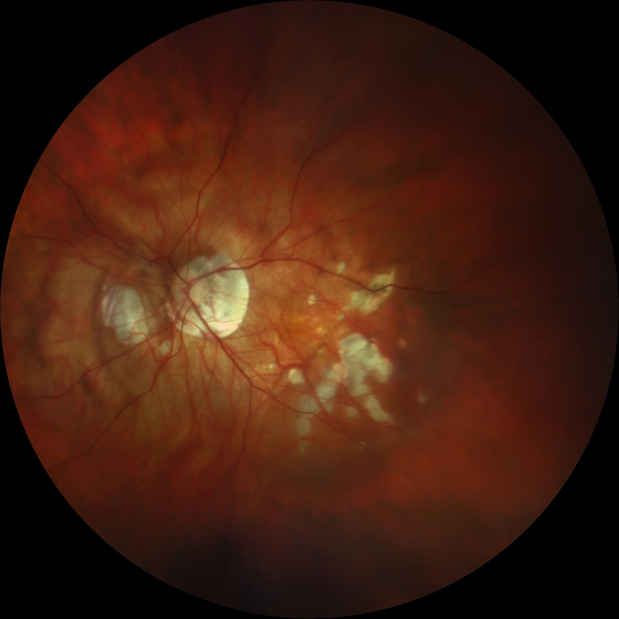
LE: Small patchy chorioretinal atrophy plaques without foveal involvement.
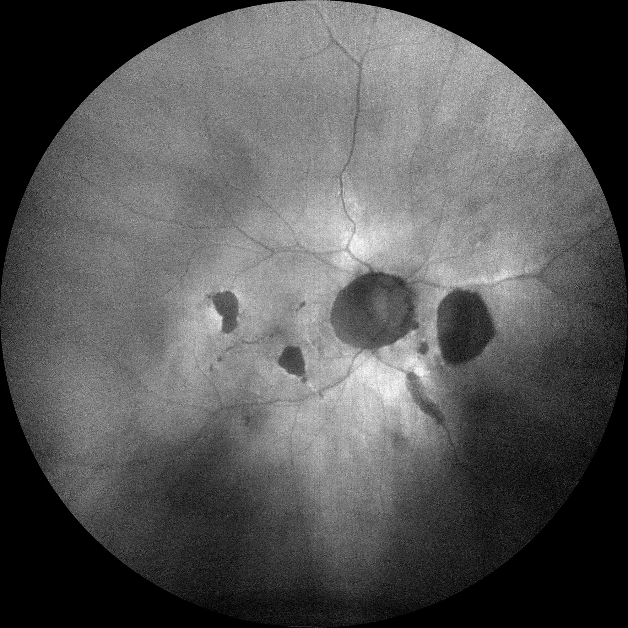
RE Autofluorescence (Clarus500, Zeiss)
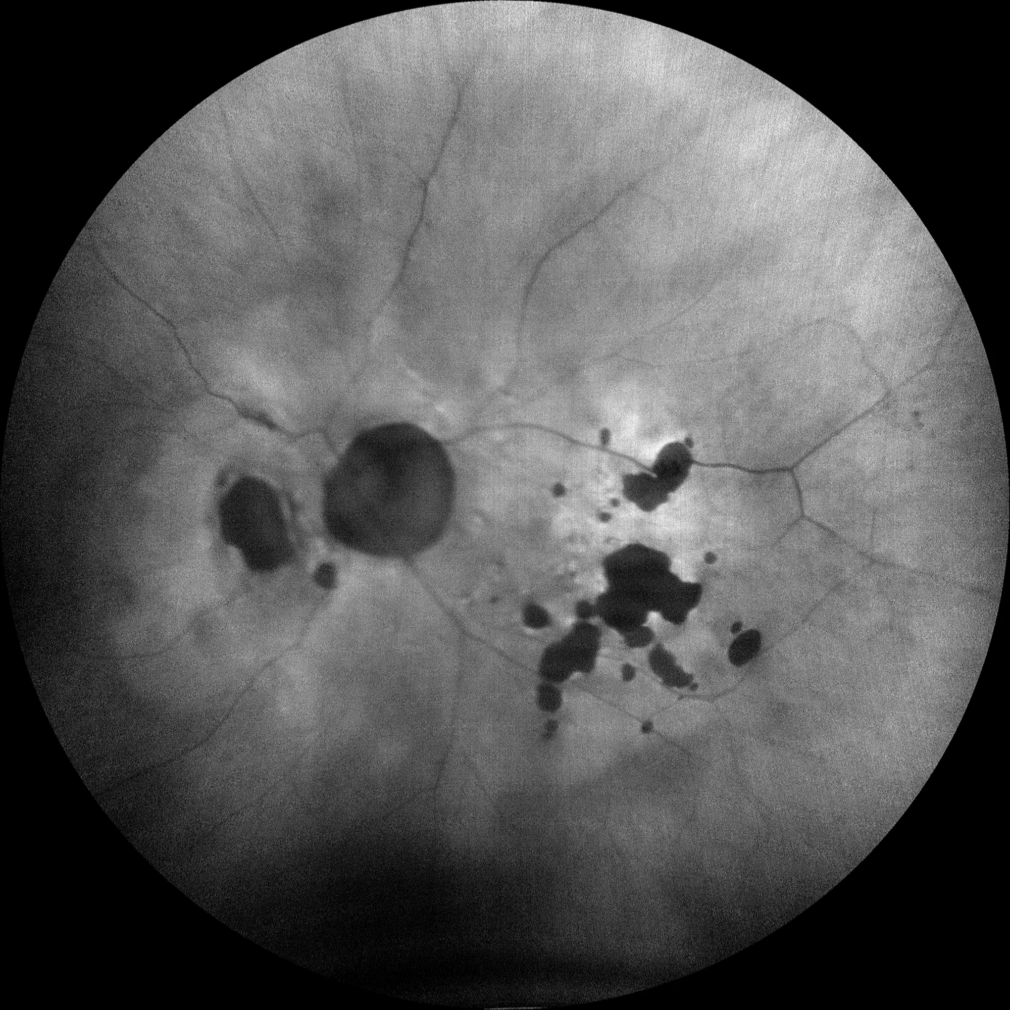
LE: Hypoautofluorescent chorioretinal atrophy plaques are notable, appearing black due to loss or absence of RPE cells.
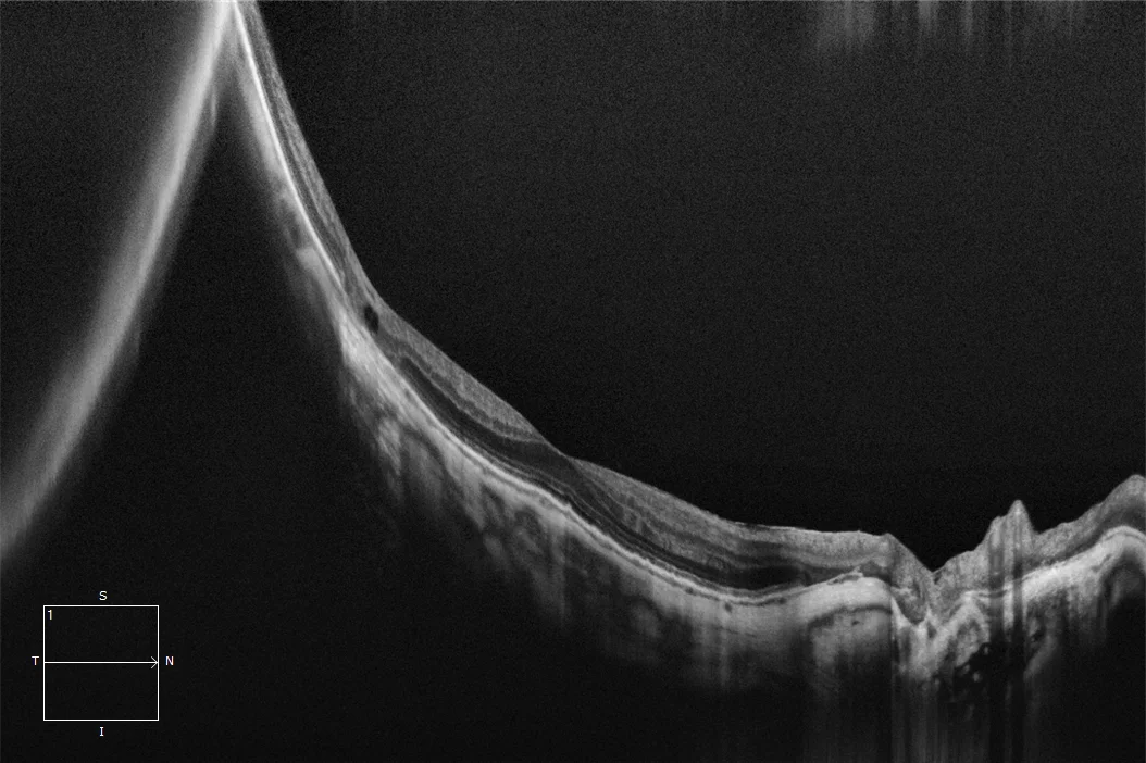
Slanted macular plane due to posterior staphyloma. An area temporal to the macula shows absence of external layers and choroid, with intraretinal cystic cavities and the inner retina directly supported on the sclera, corresponding to one of the atrophy plaques. In this section, there is no evidence of neovascular membrane.
LE: Slanted macular plane due to posterior staphyloma. No evidence of neovascular membrane.
Description
Diffuse Choroidal Atrophy. This corresponds to category 3 of the ATN classification of atrophic myopic maculopathy. It presents as one or several chorioretinal atrophy plaques in the posterior pole, manifesting as focal, circumscribed, and well-defined areas of RPE absence and loss of retinal external layers and most of the choroid. These plaques are grayish-white in color as they allow visualization of the sclera through the remaining retinal tissue. Their prevalence increases with age and axial length. Over time, the atrophy patches tend to increase in size and merge, and although not frequent, they can eventually affect the fovea. Up to 21% of cases can develop neovascular membranes associated with the atrophy patches.