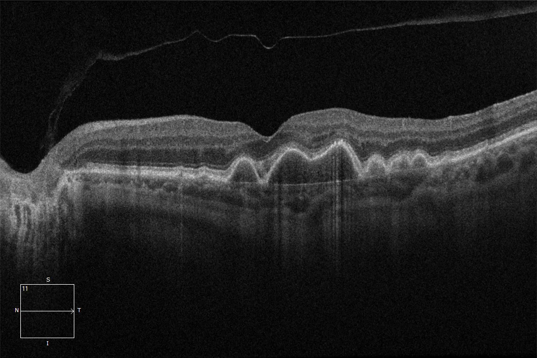
Drusenoid detachment of pigmented epithelium

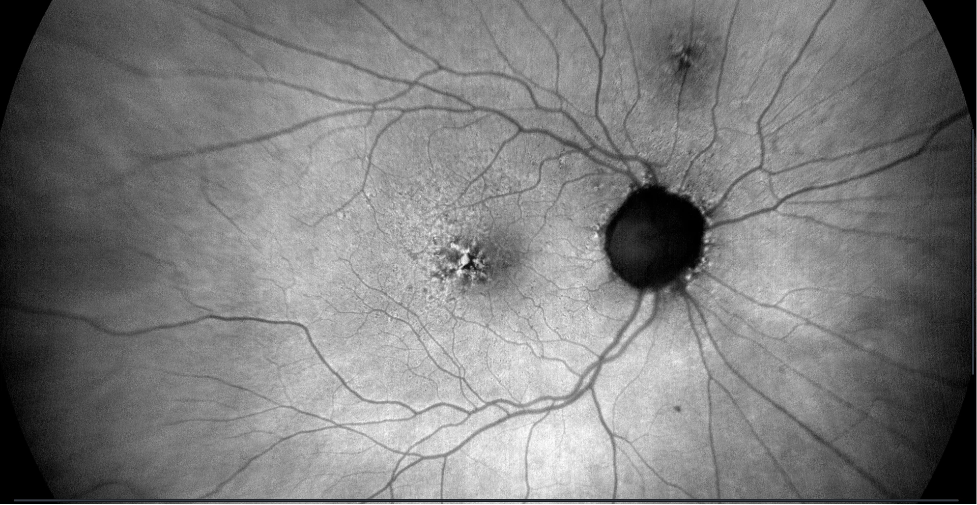
Color retinography (CLARUS 500, Zeiss): Confluent soft drusen with punctate changes in the pigmented epithelium in both eyes
OCT (Cirrus-HD 6000, Zeiss) (A): Drusenoid detachments of the pigmented epithelium in both eyes. Bruch's membrane can be clearly seen.
Autofluorescence (CLARUS 500, Zeiss): Punctate hyper- and hypoautofluorescence in both eyes, coinciding with drusenoid detachments of the pigmented epithelium

Color retinography (CLARUS 500, Zeiss): Confluent soft drusen with punctate changes in the pigmented epithelium in both eyes
Description
Drusenoid detachment of the pigmented epithelium. The presence of large soft duras causes them to merge, giving rise to an image of detachment of the pigmented epithelium with homogeneous hyperreflective material. The presence of intraretinal or submacular fluid would suggest the presence of a neovascular membrane.