Epiretinal Membrane (ERM)

Color retinography (CLARUS 500, Zeiss): Epiretinal membrane with folds at the macular level along with cuticular drusen around the lesion.
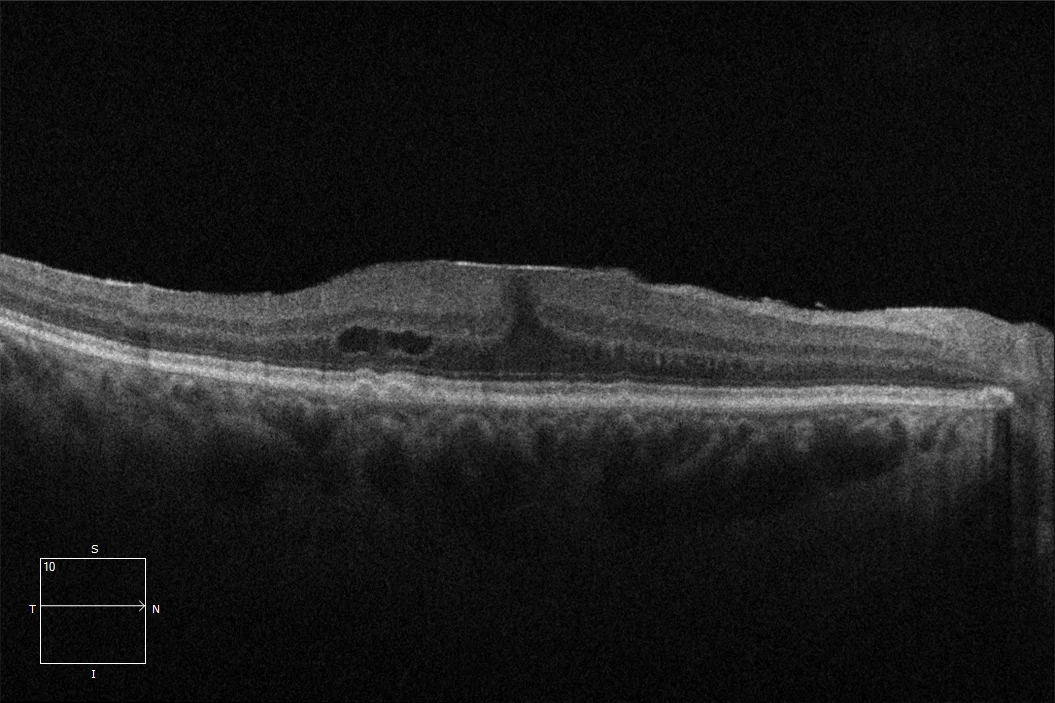
OCT (Cirrus-HD 6000, Zeiss) (A): Epiretinal membrane at the macular level that alters the foveal profile and is accompanied by macular edema and cuticular drusen.
Description
Epiretinal Membrane (ERM). It is an avascular, transparent fibrocellular tissue located on the internal surface of the retina that adheres to and covers the internal limiting membrane. It is usually located at the macular level and can be bilateral in up to 20% of cases. During examination, a shine on the retinal surface is often seen, which may be accompanied by folds. In the presented case, it is accompanied by cuticular drusen, which appear in the retinography as multiple yellowish or pale, round, and punctiform spots of 50-75 μm. Macular OCT allows us to analyze the ERM in greater detail and provides information about the prognosis, useful in making surgical decisions.