Focal choroidal excavation
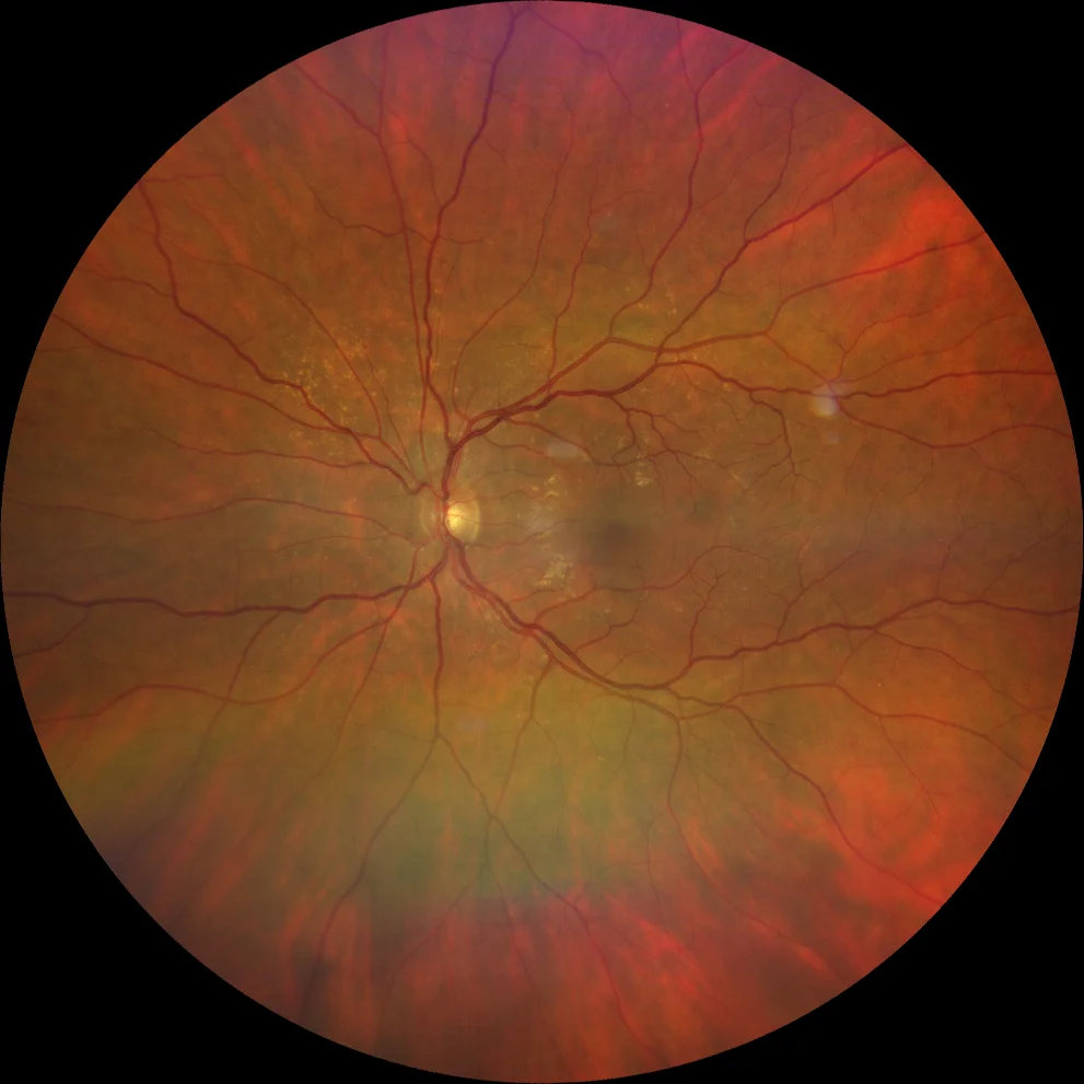
Retinography (Clarus 700, Zeiss): grayish lesion temporal to the fovea (A).
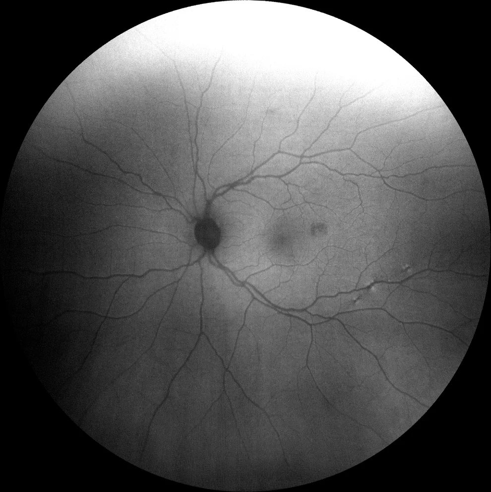
Green autofluorescence (Clarus 700, Zeiss): The lesion is best visualized in FA as a well-defined hypoautofluorescence (B).
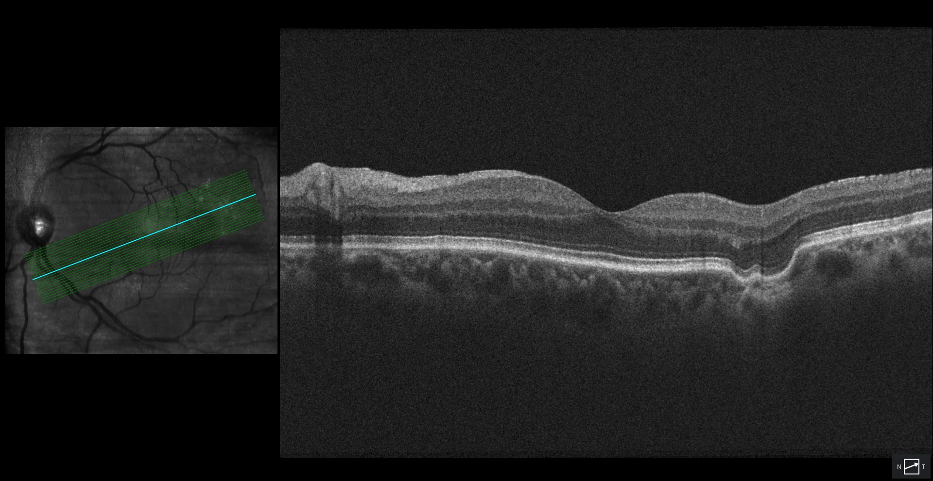
OCT (Cirrus 5000, Zeiss): RPE depression with underlying choriocapillaris atrophy. There is also partial atrophy of the large choroidal vessels at this level. The overlying retinal tissue adapts to the RPE deformation and, for this reason, focal choroidal excavation is classified as conformational (C).
Description
A 69-year-old woman comes for a check-up. She is asymptomatic.
Visual acuity 20/20 in both eyes.
The fundus shows hard drusen in the posterior pole and a subtle grayish lesion in the macula temporal to the fovea of the left eye (A). This lesion is hypoautofluorescent in green autofluorescence (B). The transverse OCT section shows a depression of the RPE with atrophy of the choriocapillaris and partially of the choroid below it. The overlying retinal tissue adapts to the depression of the RPE without leaving any hyporeflective space between them. This is a focal conformal choroidal excavation.