Focal choroidal excavation
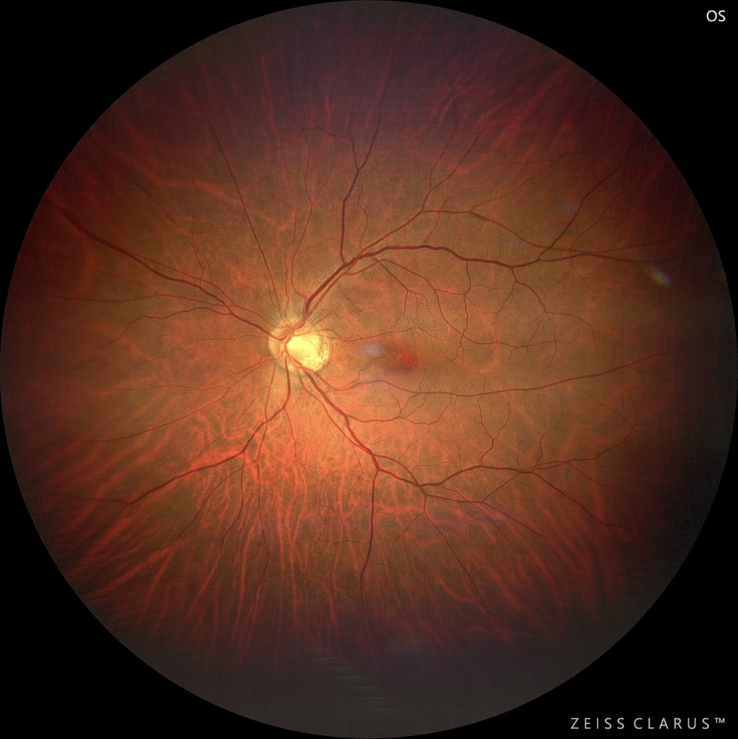
Color retinography in which we can clearly see the choroidal vessels (choroidal tessellation). Intensely reddish focus at juxtafoveal level.
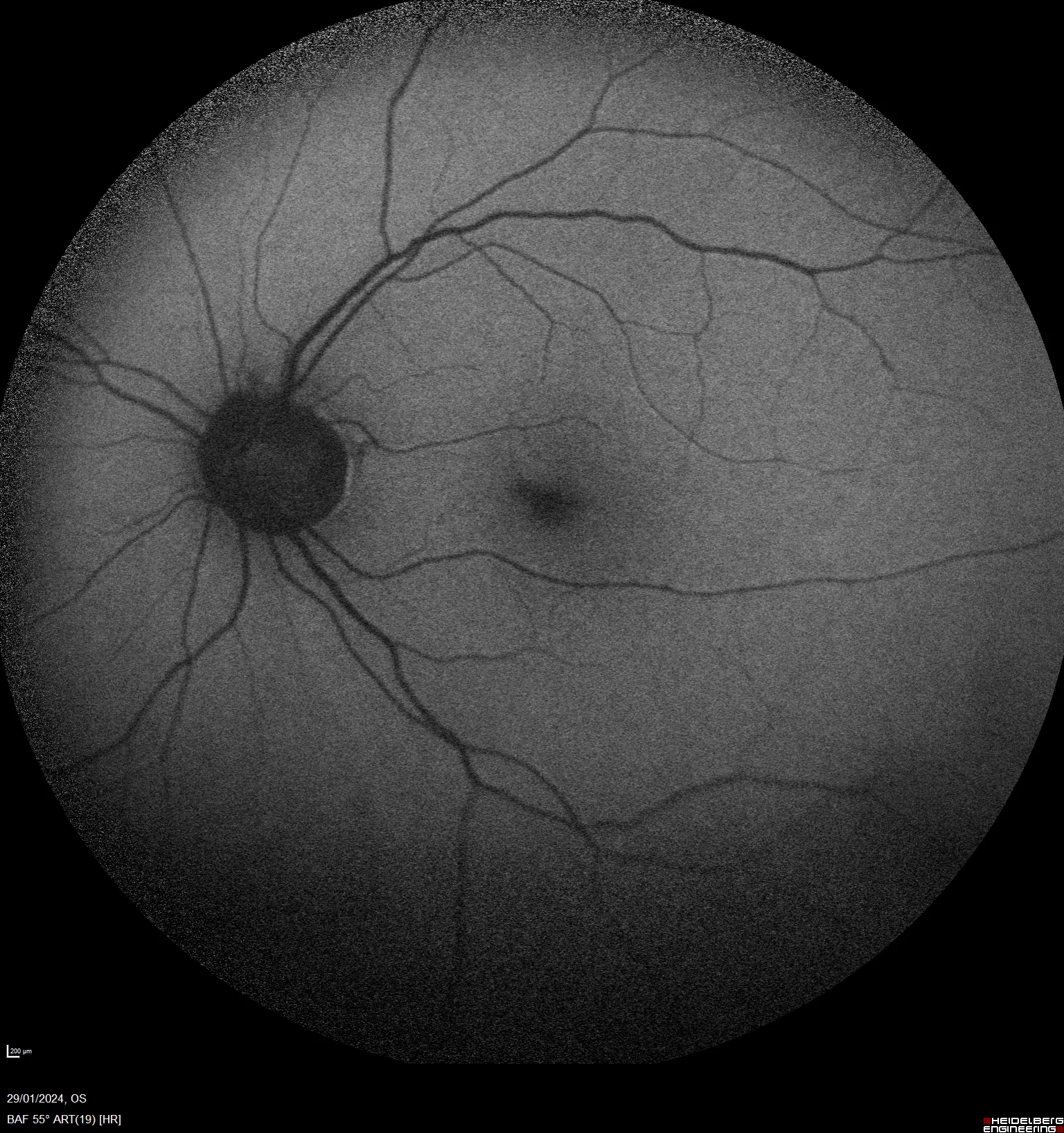
Increased intensity of macular hypoautofluorescence. No patches reflecting multifocal pigmented epithelial changes are seen.
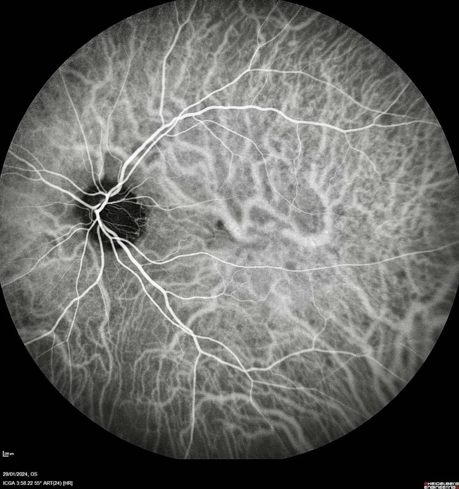
Indocyanine green shows the entry of a large-caliber choroidal vessel in the juxtafoveal region. Focal hypocyanescent defect secondary to the presence of a pigmented epithelial detachment.
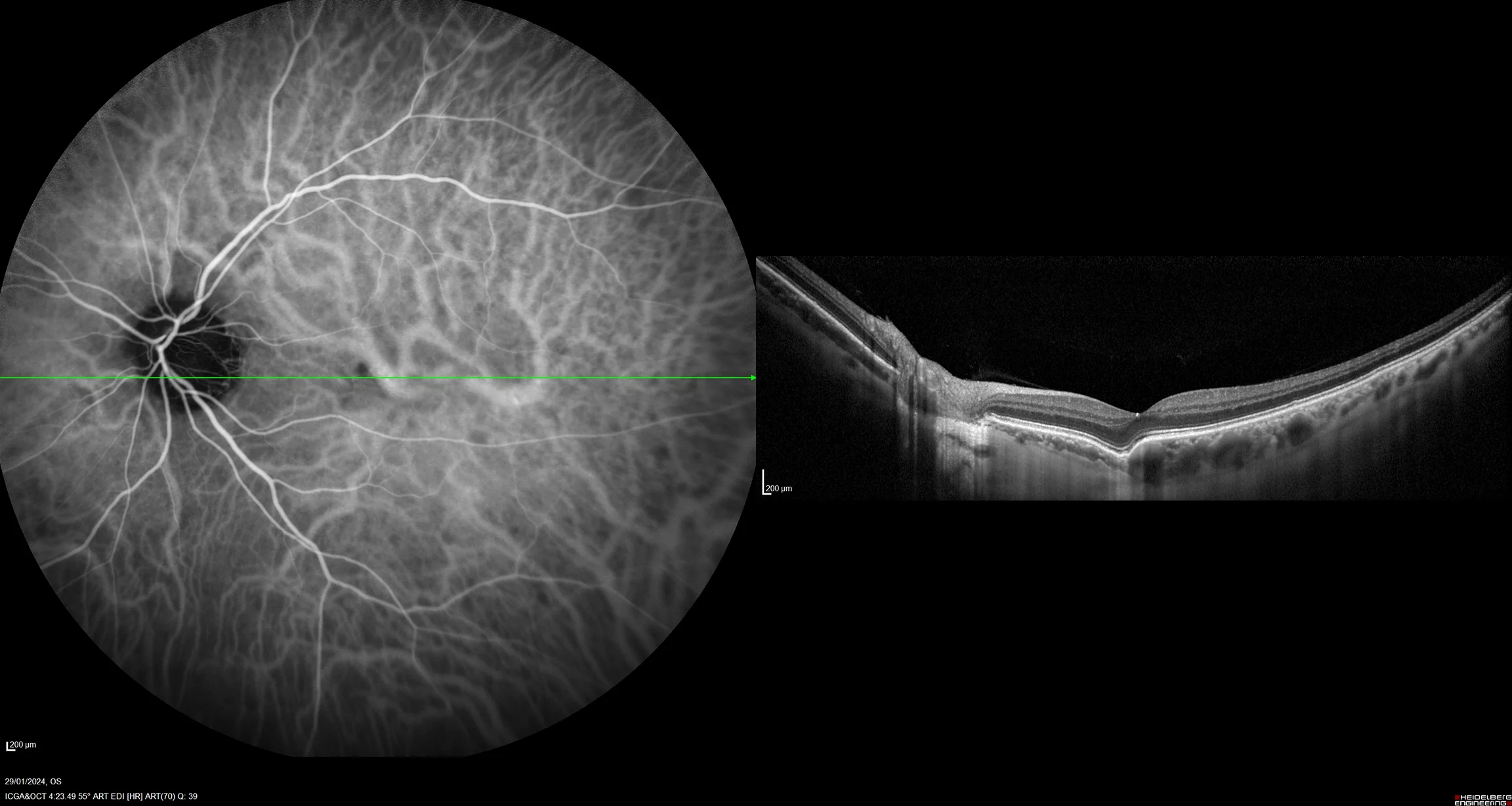
OCT shows a choroidal depression, which is accompanied by all the layers of the retina. Next to the excavation, the presence of a large choroidal vessel is notable.
Description
Focal choroidal excavation (FCE) is a rare congenital or acquired anomaly characterized by a localized depression in the choroid, observed primarily on high-resolution imaging studies such as optical coherence tomography (OCT). This condition presents with a well-circumscribed depression in the choroid, without significant alteration of the overlying retinal layers. The retina may be conformal or non-conformal, depending on whether or not it is accompanied by the pigmented epithelium and choroid.