Hypertensive retinopathy
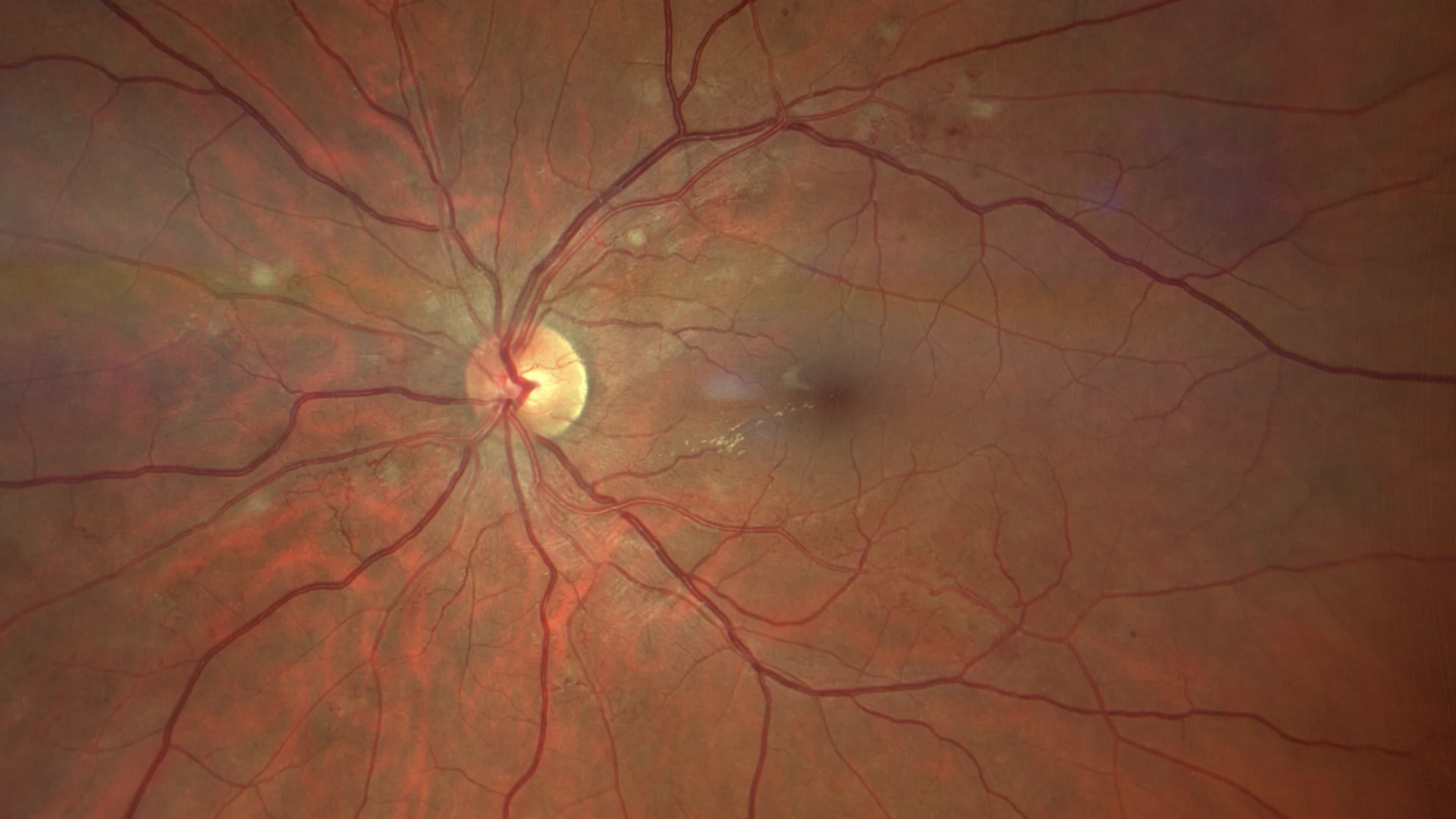
Color retinography showing both some cotton-like exudates and hard exudates, together with flame-shaped hemorrhages accompanying the nerve fibers in the posterior pole.
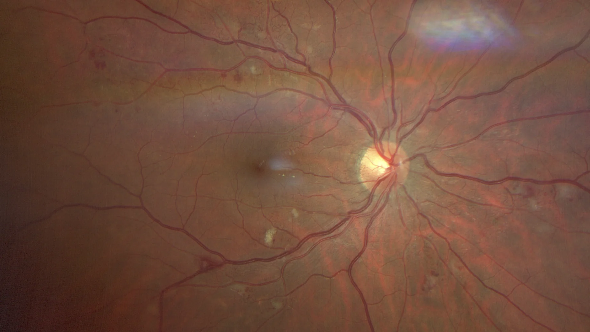
Color retinography of the olelphic eye with similar findings, and in which occlusive signs with frank arteriovenous crossings and the formation of secondary telangiectasias are evident.
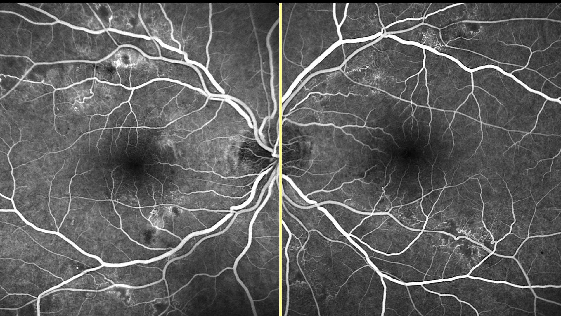
Fluorescein angiography, where capillary closures, microaneurysms, telangiectasias, and capillary leaks are observed.
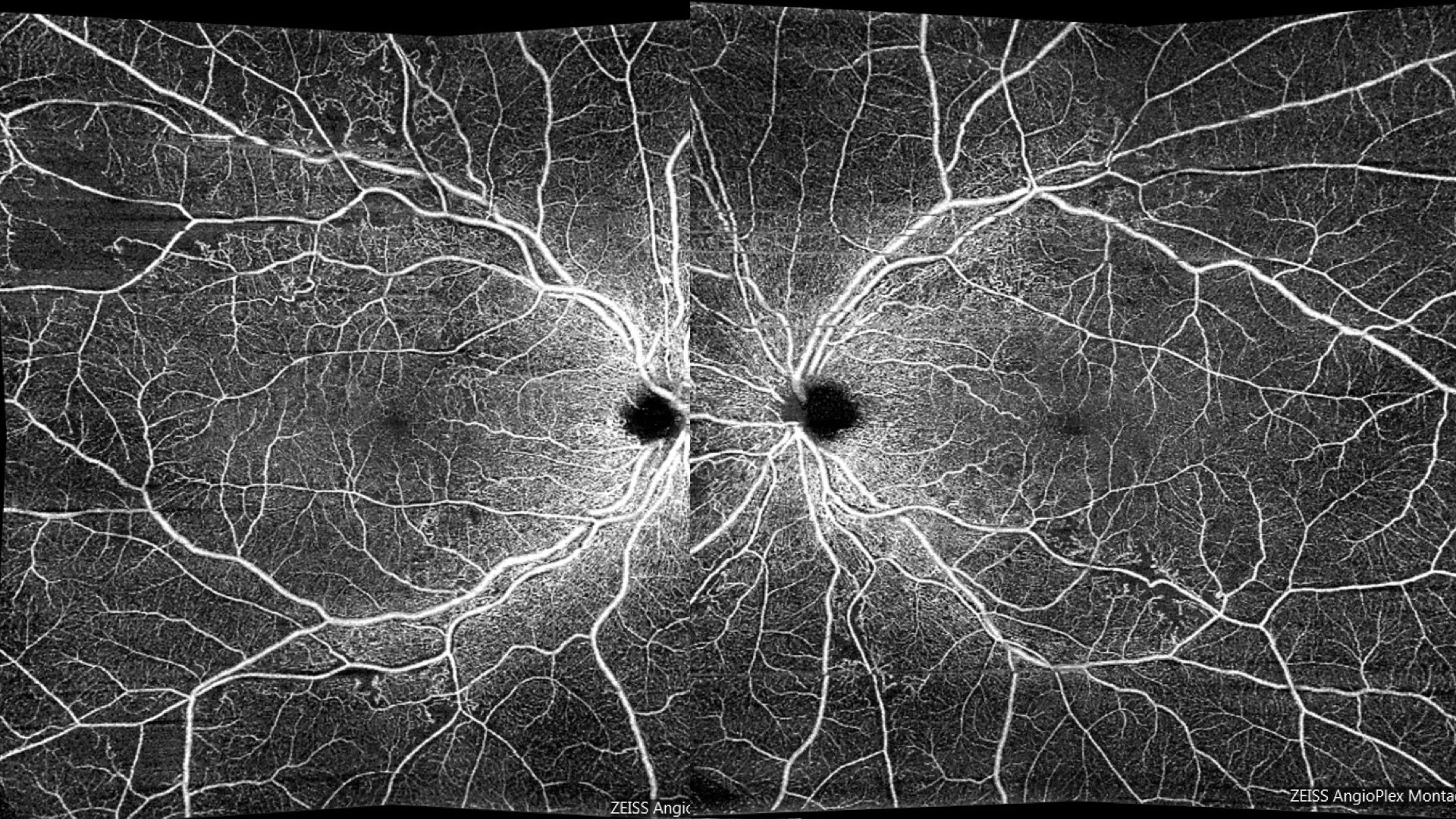
OCT angiography with Zeiss Angioplex montage showing similar vascular findings, with special demarcation of ischemia in the capillary plexus of the superior temporal vascular arcade of the RE.
Description
Hypertensive retinopathy is an ocular manifestation of systemic hypertensive disease, characterized by vascular changes in the retina resulting from chronically elevated blood pressure. On fundusography, typical findings include generalized narrowing of the retinal arterioles, changes in the arteriovenous relationship, pathological arteriovenous crossings (Gunn’s sign), and in more advanced stages, retinal hemorrhages, cotton-wool spots, and papilledema.
These changes are commonly classified according to the Keith-Wagener-Barker scale. Management of hypertensive retinopathy involves strict blood pressure control to prevent progression of retinal damage and mitigate the risk of associated complications such as macular edema and ischemic optic neuropathy.