Idiopathic choroidal folds
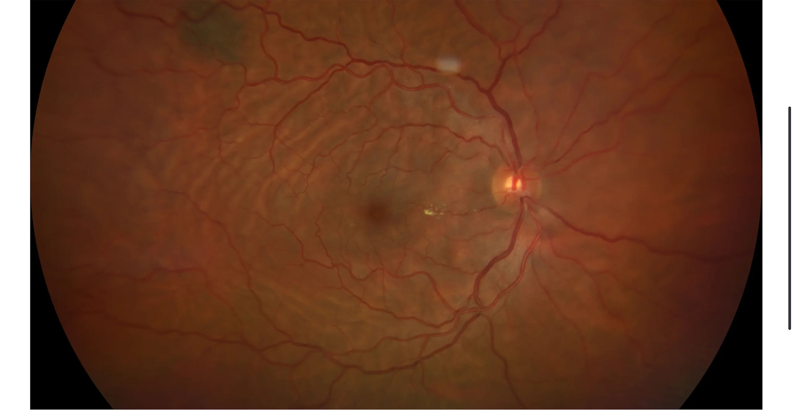
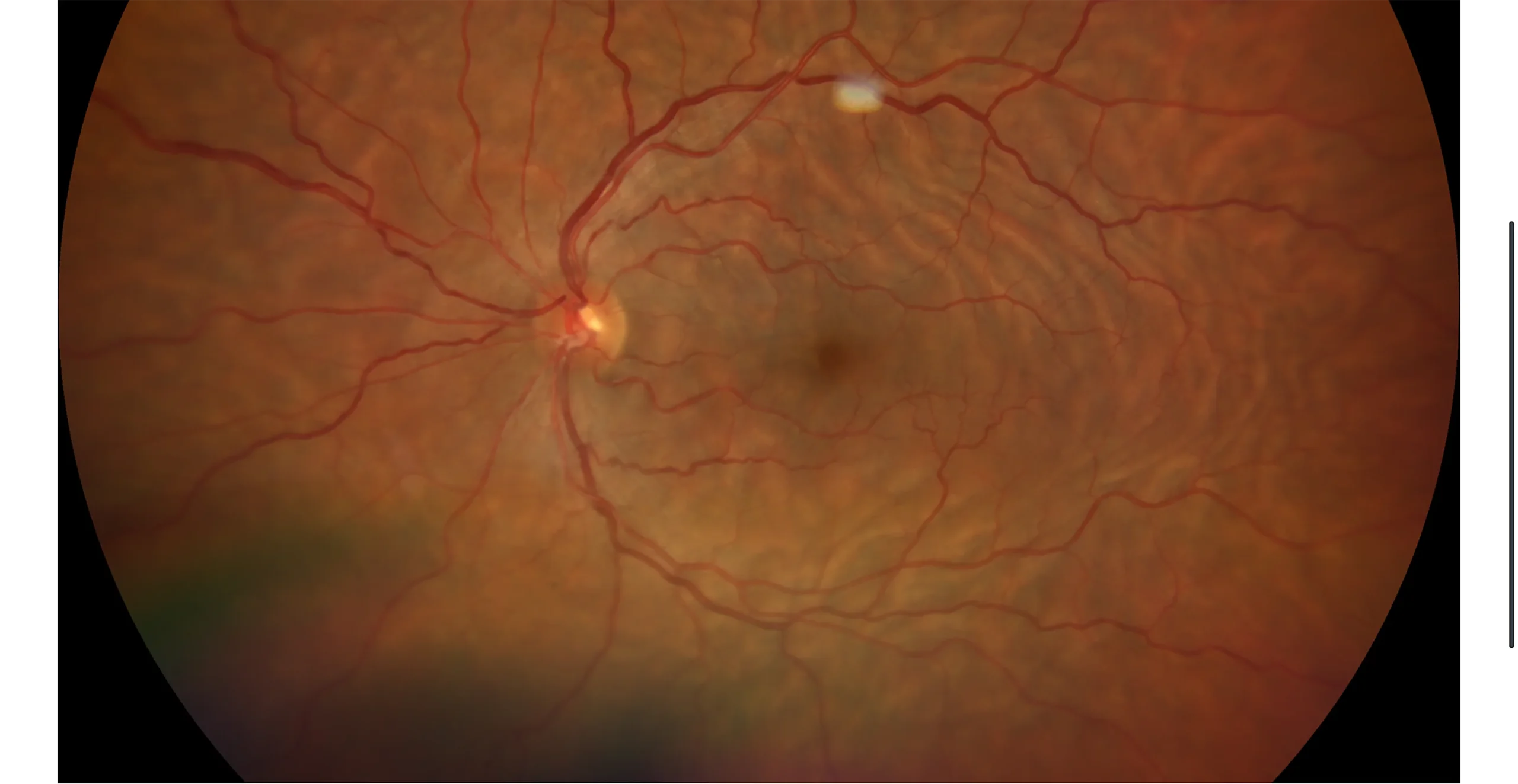
Retinography: Choroidal folds with a circular arrangement following the path of the vascular arcades
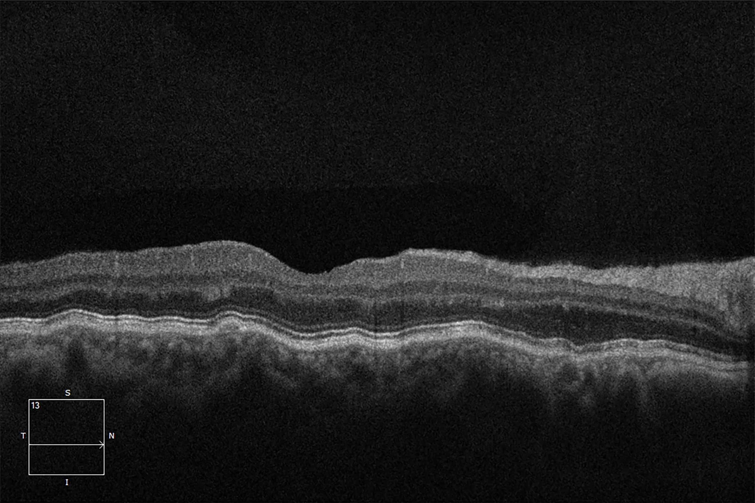
OCT OD
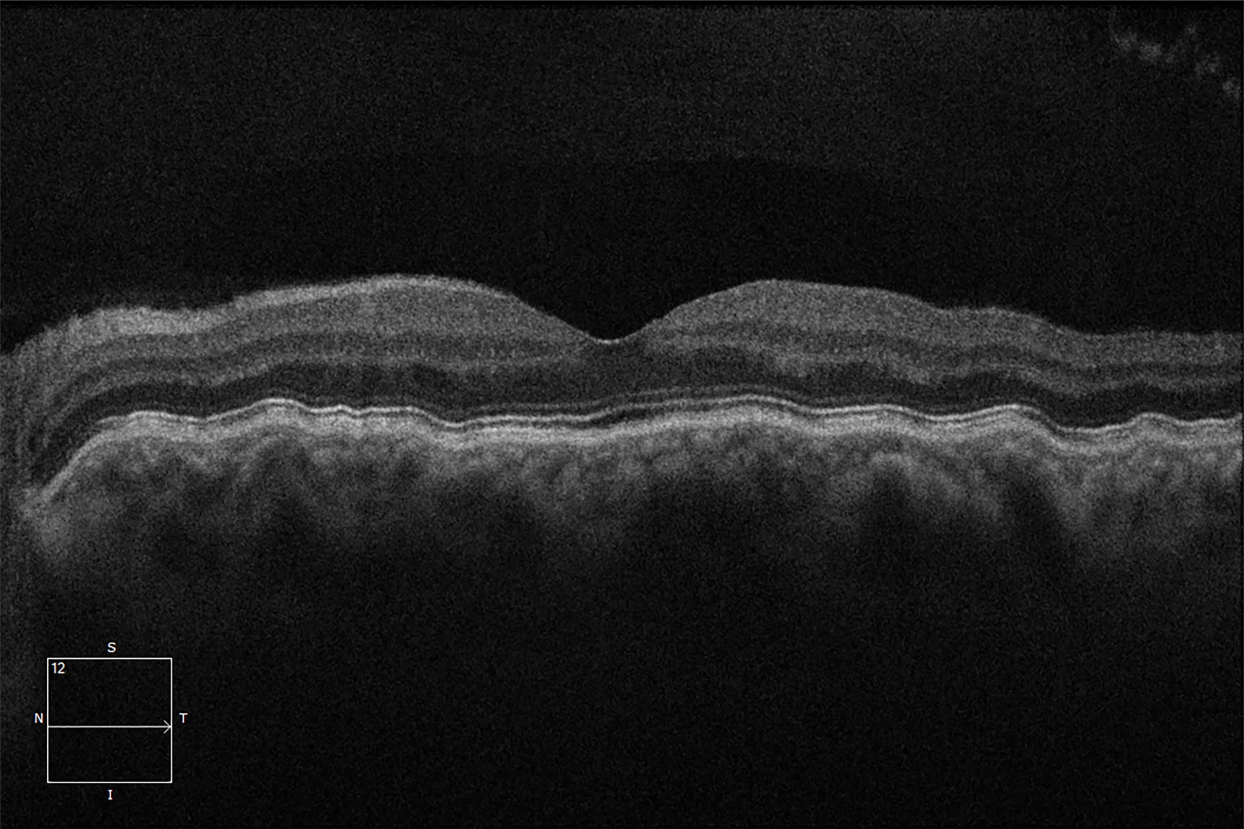
OCT OS
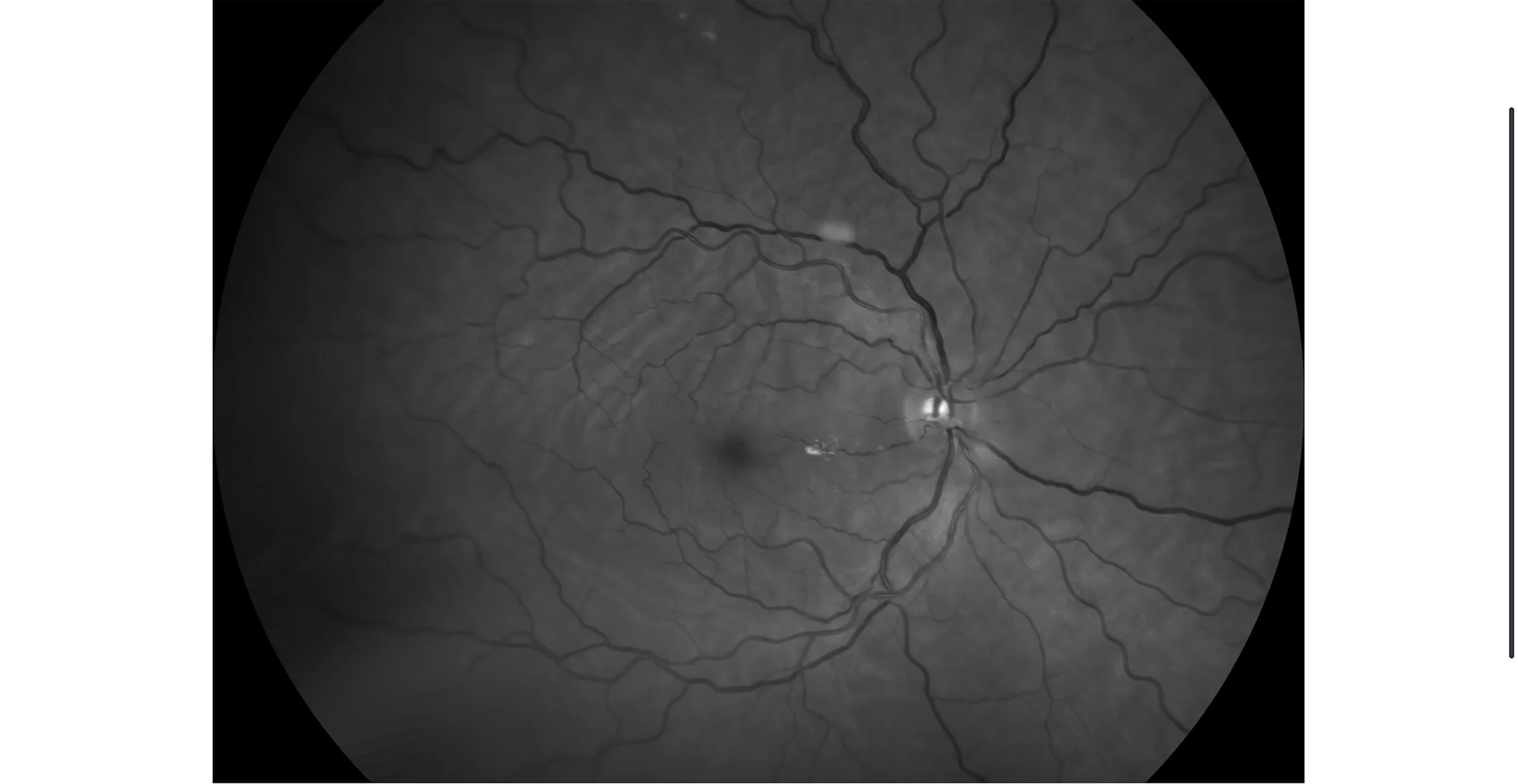
OD Retinography with red-free filter: Visible idiopathic choroidal folds
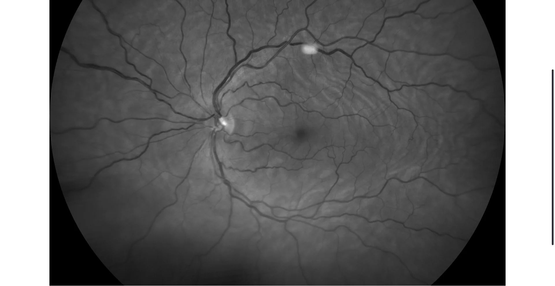
OS Retinography with red-free filter: Visible idiopathic choroidal folds
Description
Idiopathic choroidal folds. It is an entity with a low prevalence in the general population. It involves undulations of the choroid, Bruch’s membrane, the pigmented epithelium, and the outer retinal layers. In the case of idiopathic folds, the diagnosis is usually made as an incidental finding and patients are asymptomatic. They can also be observed in cases secondary to orbital tumors, hypotony, papillitis, hyperopia, posterior scleritis, and previous scleral surgery. They should be differentiated from retinal folds, characteristically present in alterations of the retinal surface.
Comments
- Retinography (CLARUS 500, Zeiss): Choroidal folds following the direction of the vascular arcades. In the right eye, it presents lipid exudates secondary to the underlying diabetic retinopathy.
- OCT (Cirrus-HD 6000, Zeiss): Choroidal folds distorting the macular structure. They are limited to the choroid, pigmented epithelium, and outer layers.