< back
Idiopathic macular hole (stage 3)
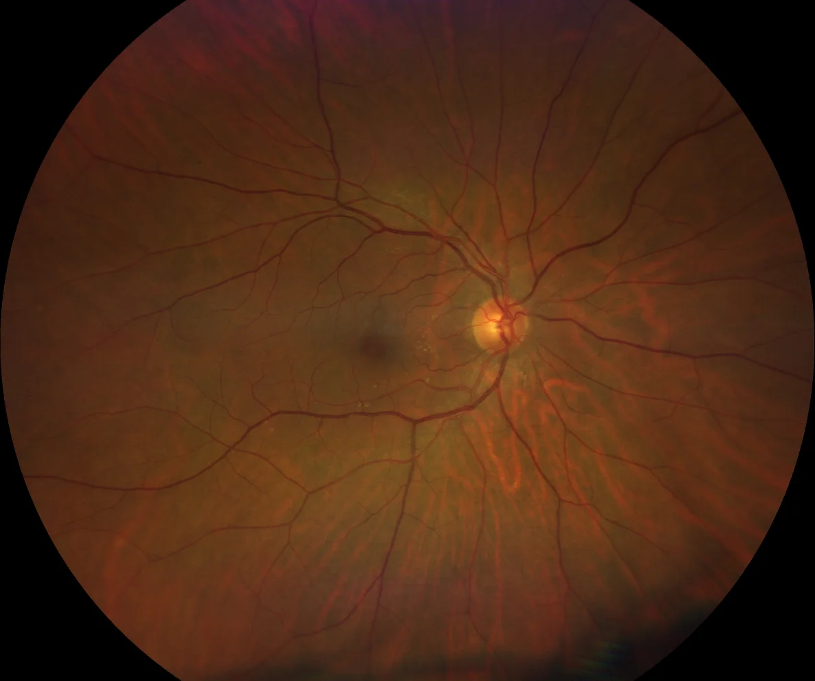
Color retinography (CLARUS 500, Zeiss): Macular hole with small prefoveal operculum.
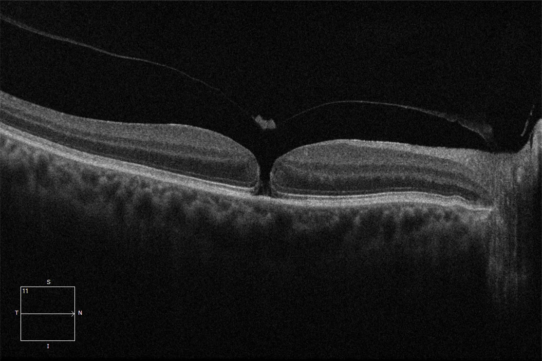
OCT (Cirrus-HD 6000, Zeiss) (A): Stage 3 macular hole with the posterior hyaloid partially detached, evident in the prefoveal plane and with an adherent operculum.
Description
This is a discontinuity of the foveal area secondary to vitreomacular traction and, subsequently, to progressive tangential traction. In this case, the posterior hyaloid is shown detached in the foveal plane, constituting stage 3.