Idiopathic macular telangiectasias type 1 (MacTel1)
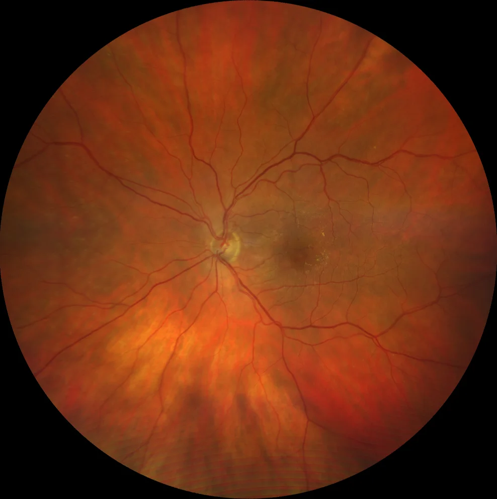
Retinography (Clarus 700, Zeiss): hard macular exudates, especially temporal to the fovea. Reddish, punctate perifoveal lesions (corresponding to MacTel1). A rounded, yellowish lesion corresponding to a saccular vascular dilatation is also observed (A).
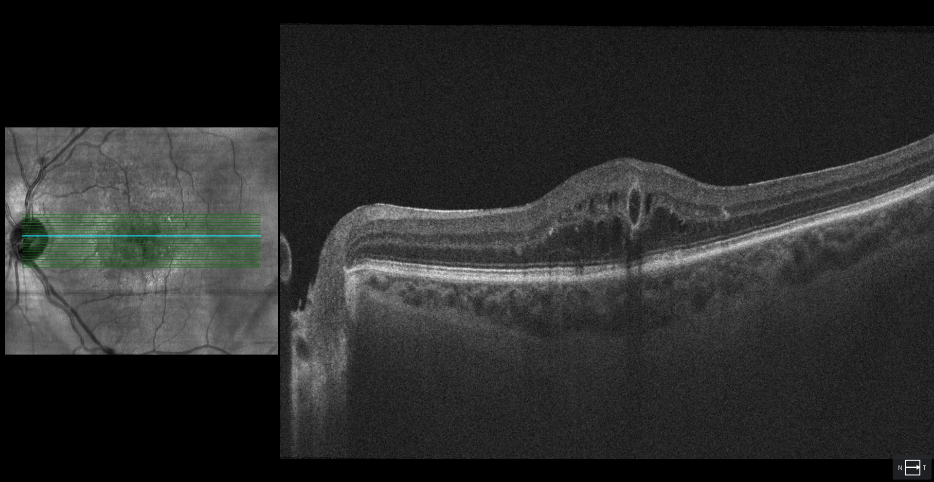
OCT (Cirrus 5000, Zeiss): intraretinal fluid, both in the inner nuclear layer and in the outer nuclear layer. Several rounded lesions with hyporeflective content and hyperreflective walls are also observed (one of them shown) that correspond to telangiectatic dilatations of the perifoveal capillaries (B).
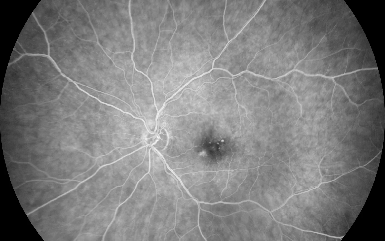
AGF (Clarus 700, Zeiss): hyperfluorescent lesions in early stages (C1) with contrast leakage in late stages (C2) corresponding to saccular dilatations of the perifoveal capillaries.
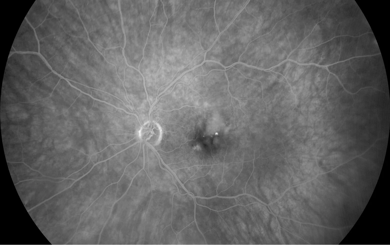
AGF (Clarus 700, Zeiss): hyperfluorescent lesions in early stages (C1) with contrast leakage in late stages (C2) corresponding to saccular dilatations of the perifoveal capillaries.
Description
An 81-year-old pseudophakic woman presented with vision loss in her left eye. She had no history of diabetes mellitus.
In the fundus of the left eye, hard macular exudates accompanied by reddish punctate lesions were observed. Intraretinal fluid was observed on OCT, as well as rounded lesions with hyporeflective content and hyperreflective walls. The FAG showed saccular dilatations of the perifoveal capillaries in early stages, with contrast leakage in late stages. No peripheral vascular dilatations were observed and the right eye was normal. A diagnosis of idiopathic macular telangiectasias type 1 or MacTel1 was made. Treatment with anti-VEGF drugs was proposed. After 1 injection of aflibercept and 1 injection of brolucizumab and given the absolute lack of response to them, it was decided not to treat further.