Juvenile Glaucoma
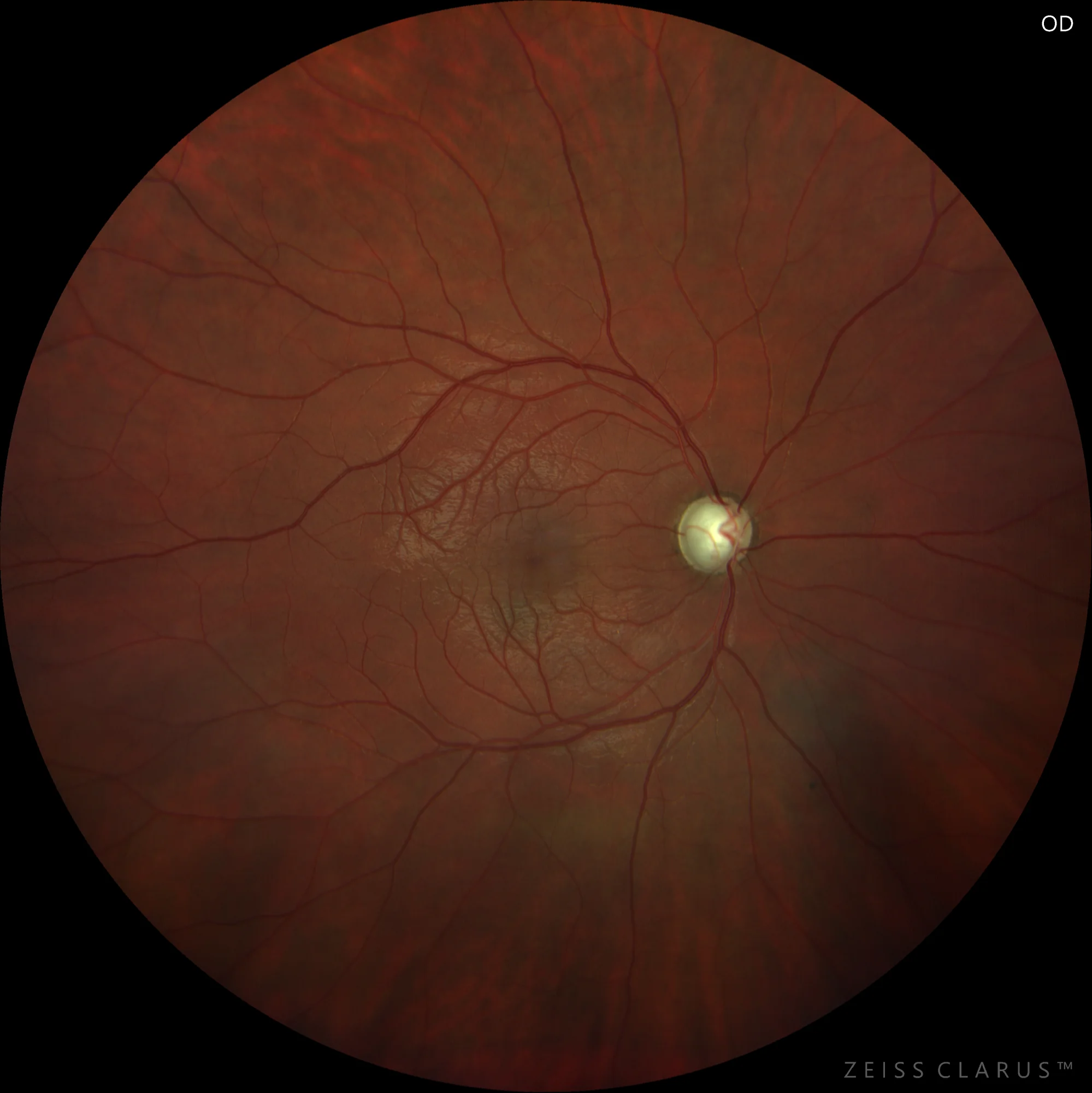
Figure 1. Color retinography of the right eye showing optic disc with marked decrease in neuroretinal rim 360º and cup-to-disc ratio of 0.9.
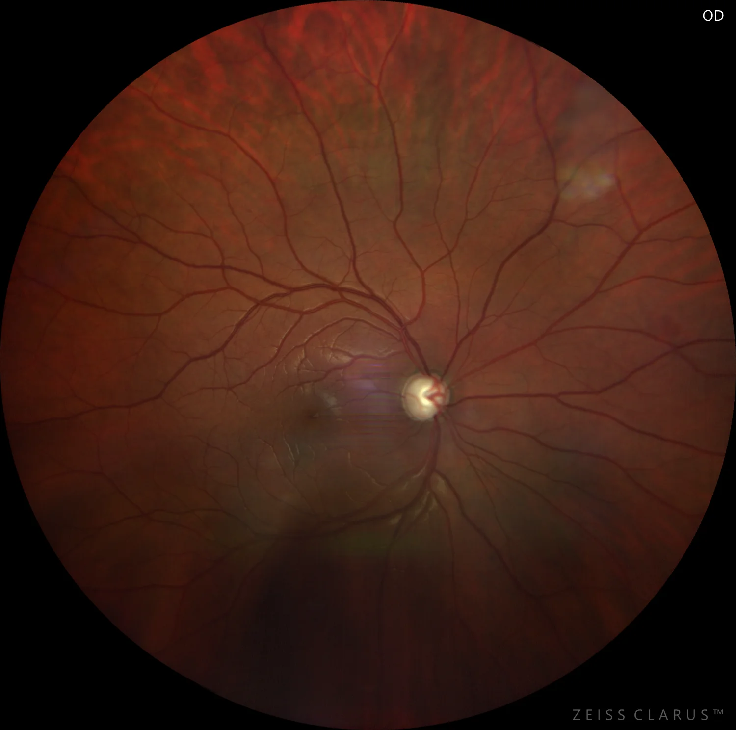
Figure 2. Color retinography of the right eye 1 year after diagnosis, with optic disc pallor and complete cupping.
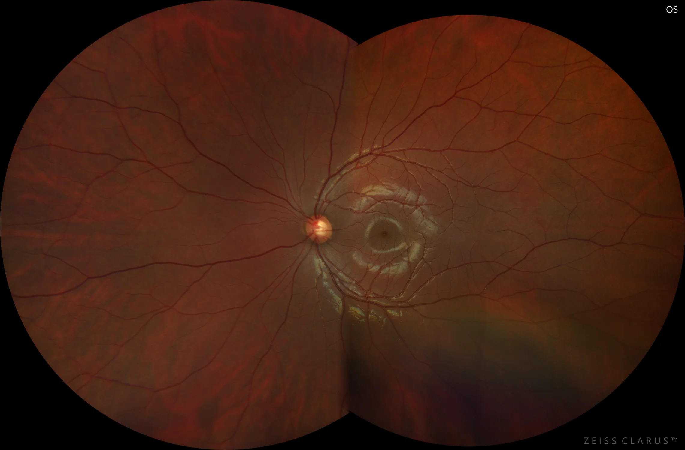
Figure 3. Color retinography of the left eye at the time of onset, with cup-to-disc ratio of 0.4 and good neuroretinal rim.
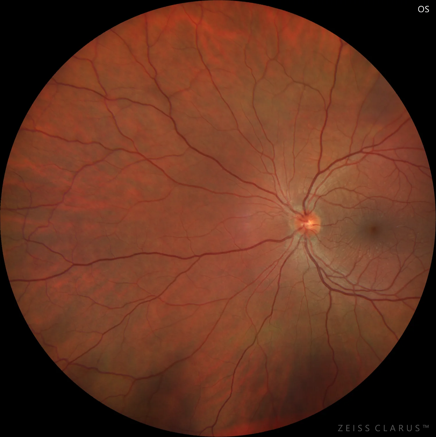
Figure 4. Color retinography of the left eye 1 year after diagnosis, with vertical elongation of the optic disc and preserved neuroretinal rim.
Description
Juvenile glaucoma is a primary open-angle glaucoma with hereditary characteristics in certain subtypes that appears at early ages. Unlike congenital glaucoma, which presents with buphthalmos, megalocornea, and Haab’s striae, we do not find alterations in the anterior pole. Gonioscopy reveals an open angle without dysgenetic alterations.
The diagnosis is clinical, observing high intraocular pressures, increased optic disc cupping with focal or generalized thinning of the neuroretinal rim, and visual field defects. Genetic testing helps us locate the mutation, with alterations in chromosome 1q being characteristic in autosomal dominant juvenile glaucoma.
Juvenile glaucoma presents a faster and more severe progression than adult primary open-angle glaucoma, debuting with higher intraocular pressures that respond poorly to topical treatment, often requiring earlier surgical intervention.