Macula in dome
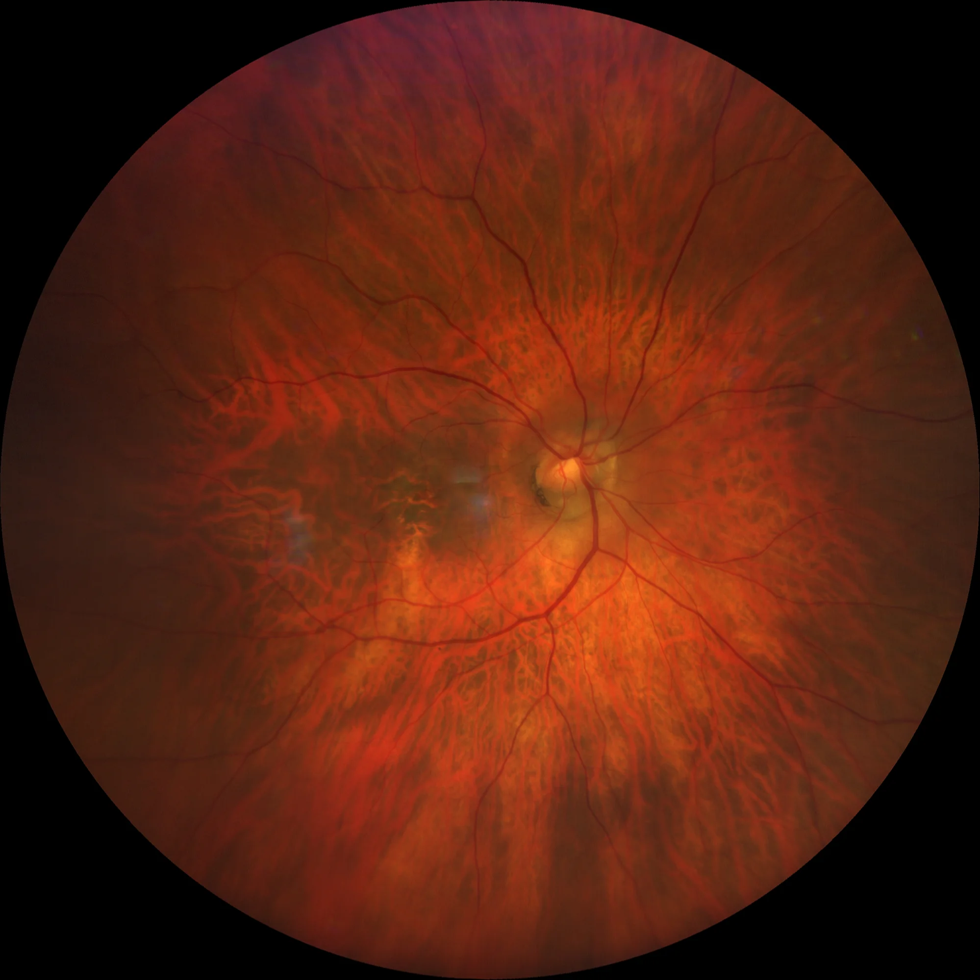
Retinography OD: Tessellated fundus. Somewhat oblique implantation papilla with peripapillary intrachoroidal cavitation. Macular pigmentary alteration with transparency of choroidal vessels.
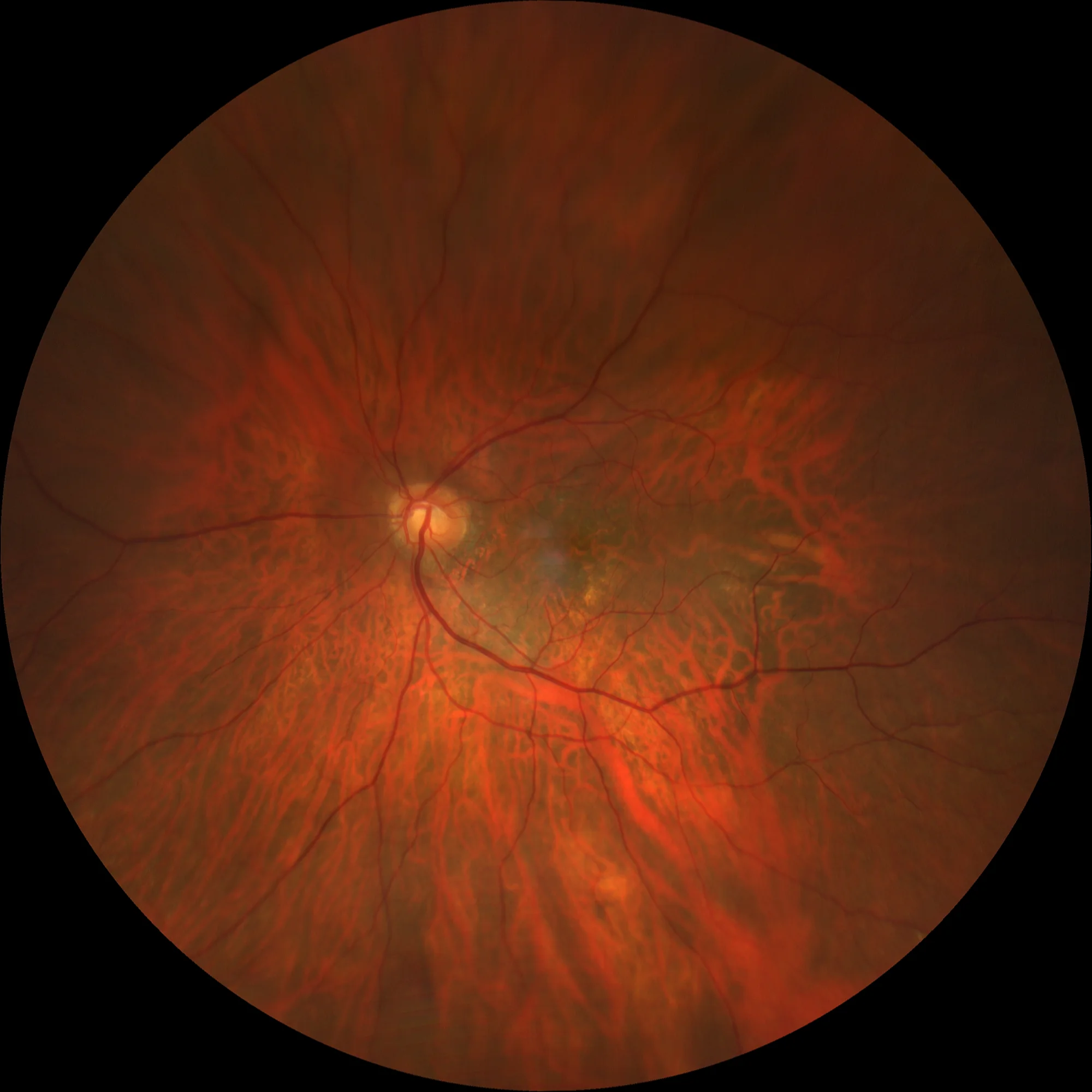
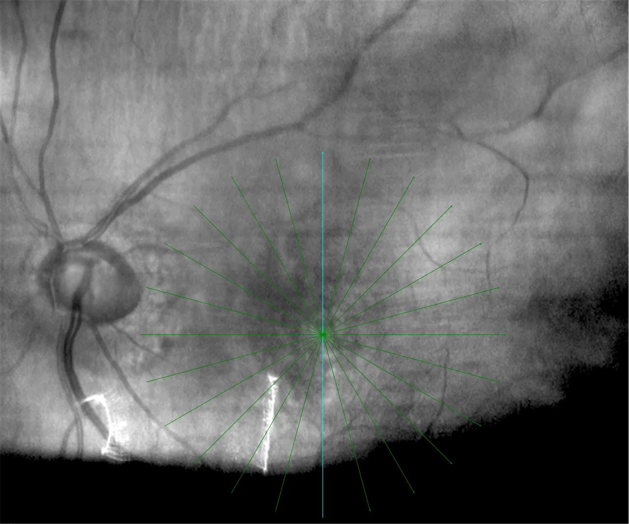
Left retinal image: Tessellated fundus. Implantation papilla somewhat oblique. Minimal macular pigmentation alteration.
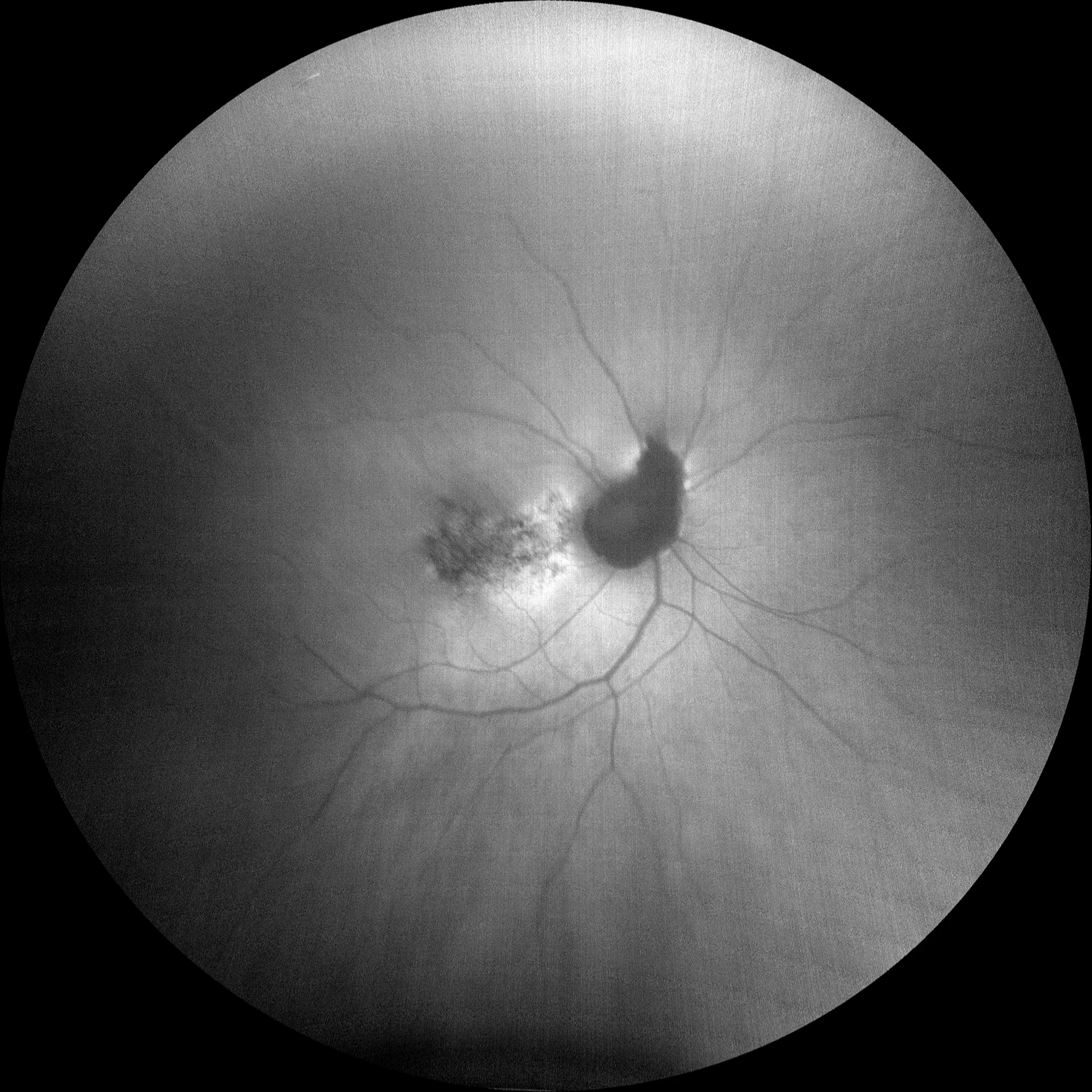
OD autofluorescence: Macular pigmentary alteration that is reflected in autofluorescence as hypo and hyper autofluorescent punctates.
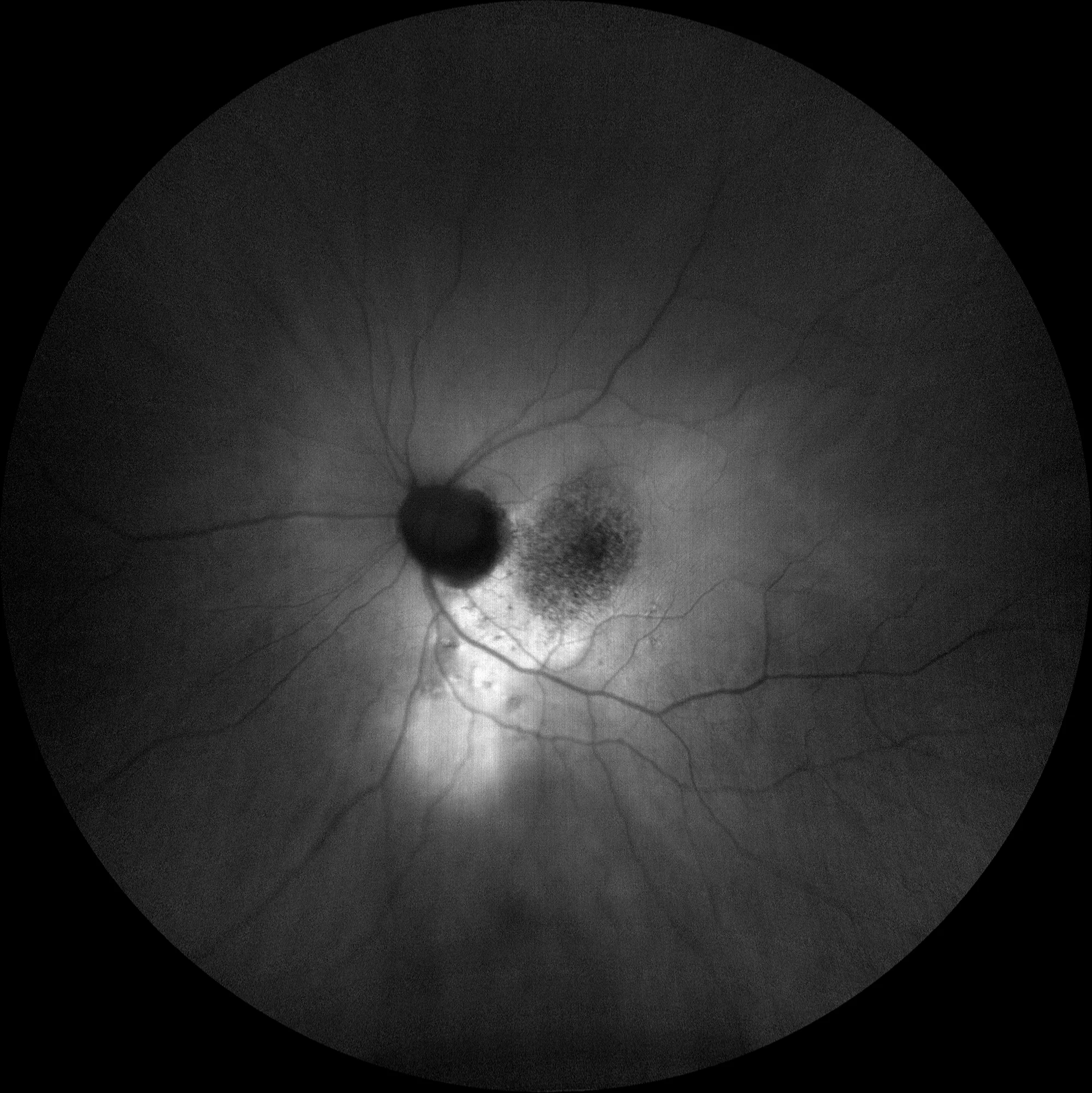
LE autofluorescence: Hypoautofluorescent macular pigmentary alteration with a hyperautofluorescent trailing pattern towards the bottom similar to that found in central sera chorioretinopathy and which seems to reflect the presence of RSF.
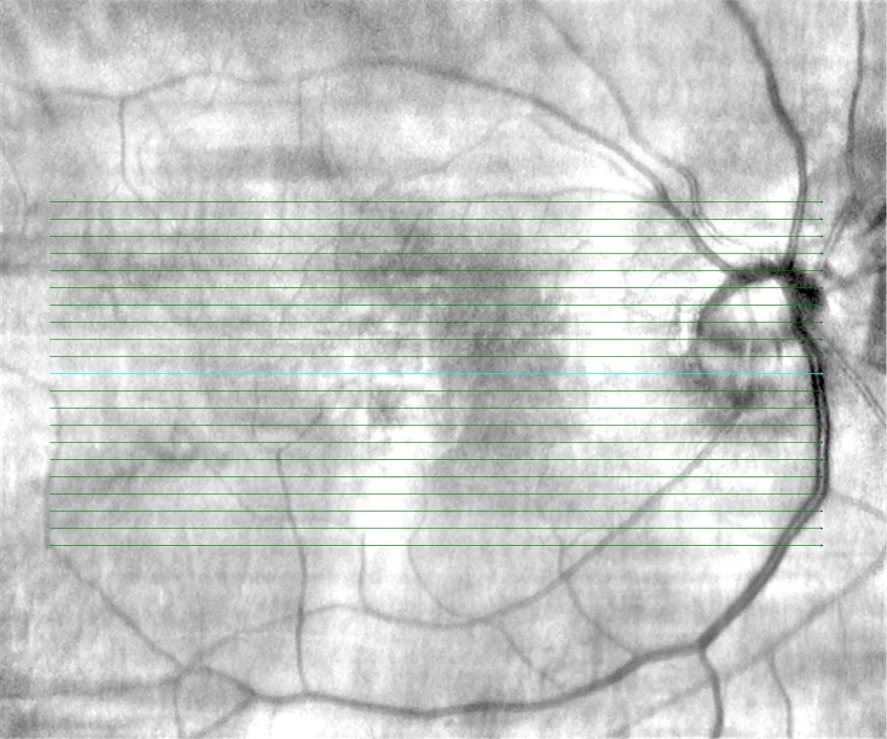
Horizontal macular OCT OD: In this slice we can see the dome-shaped macula. Greater choroidal thickening can be seen at the apex of the dome compared to the edges, which some authors have related to the formation of RSF. The existence of a peripapillary intrachoroidal cavitation can also be seen as an intrachoroidal hyporeflective cavity adjacent to the papilla.
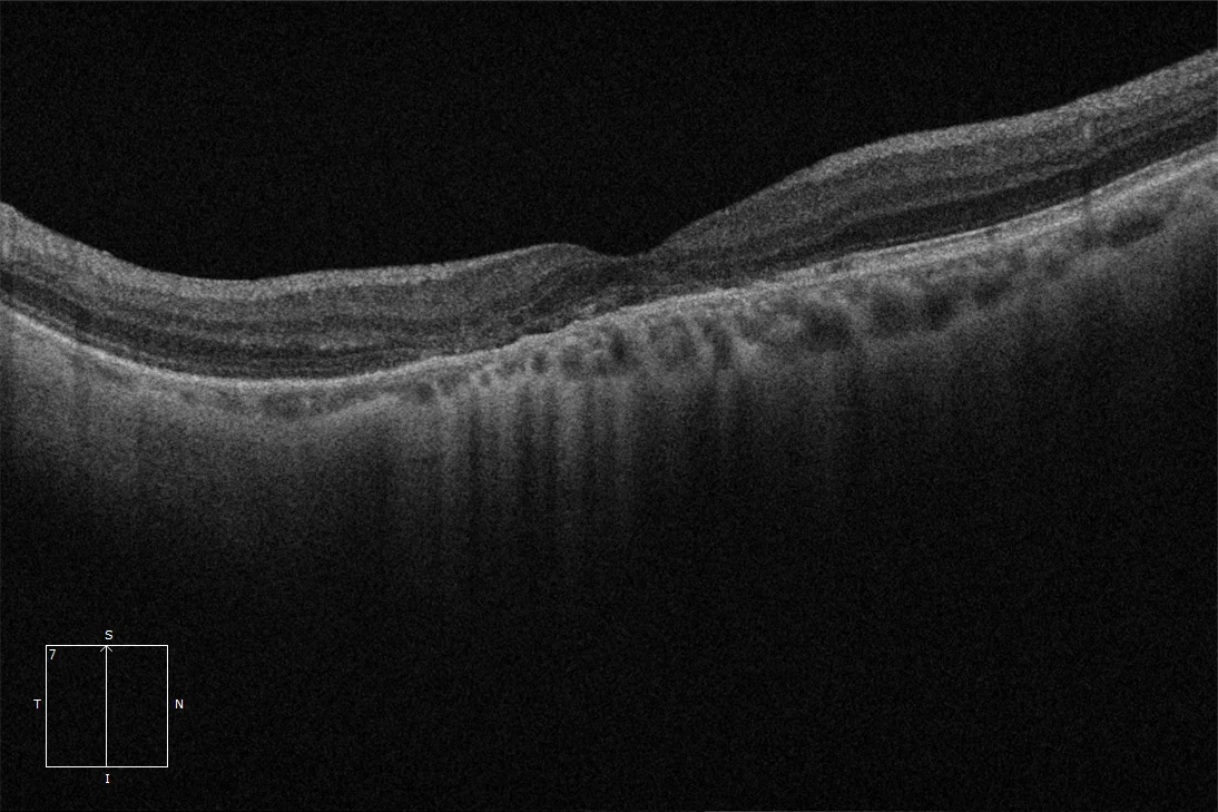
Vertical macular OCT OD: In this section the macula is flat because it is a section parallel to the dome, which in this case is oval vertically. An alteration and irregularity of the retinal pigment epithelium (RPE) and external layers can be seen.
Horizontal macular OCT LE: Dome-shaped macula with FSR. Choroidal thickening at the apex of the dome is also observed, as well as RPE irregularity.
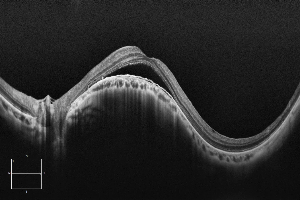
Vertical macular OCT LE: In this section the macula appears flat for the reasons already mentioned, and may simulate a central serous chorioretinopathy. As in the previous section, we observe a thinned choroid but thickened in the fovea and an irregularity of the RPE.
Description
Gaucher defined it as a convex protrusion within a posterior staphyloma, although it is currently considered a new form of staphyloma. Its pathogenesis seems to involve the existence of a scleral thickening under the macula compared to the edges of the dome where the sclera is thinned, which could explain why the thickened sclera area protrudes and causes a convex bulge of the macula. Both the dome macula and the staphyloma rim (macula located at the edge of a staphyloma) are susceptible to producing common and specific pathology, among which the appearance of subretinal fluid (SRF) or macular neurosensory detachments with alteration of the retinal pigment epithelium (RPE) stands out, for which there is no effective treatment. The subretinal fluid can vary in quantity and even remit spontaneously, and although vision usually remains stable for years, in the long term it can cause visual impairment.