Macular hemorrhage
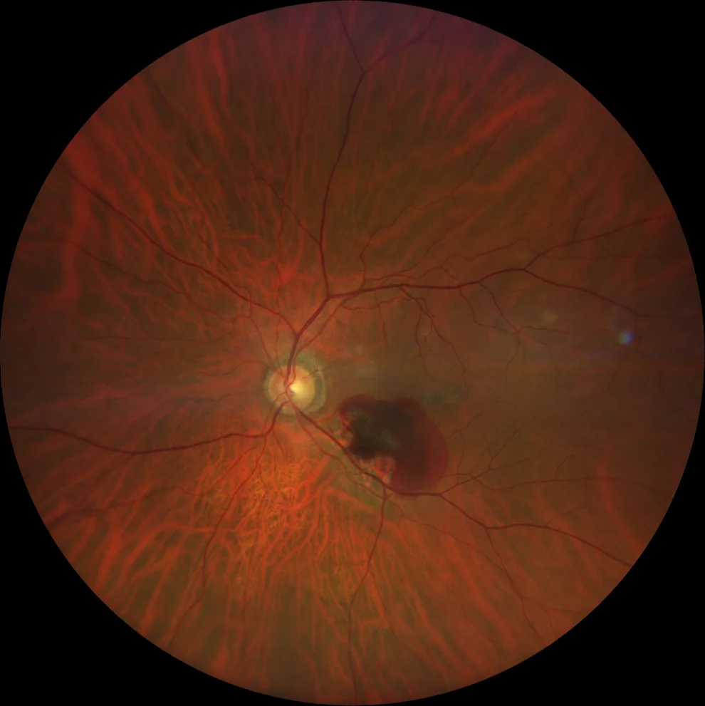
A: Macular hemorrhage with foveal involvement. The hemorrhage has different intensities, suggesting blood at different levels.
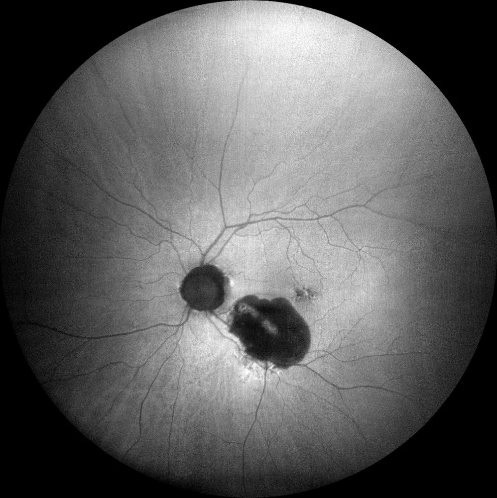
B: the lesion is hypoAF due to the screening effect of the hemorrhage that does not allow the baseline AF of the RPE to be seen.
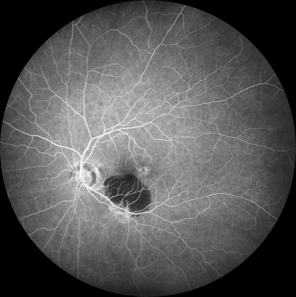
C: the lesion is hypofluorescent due to the screening effect of the hemorrhage, which does not allow the background choroidal fluorescence to be seen.
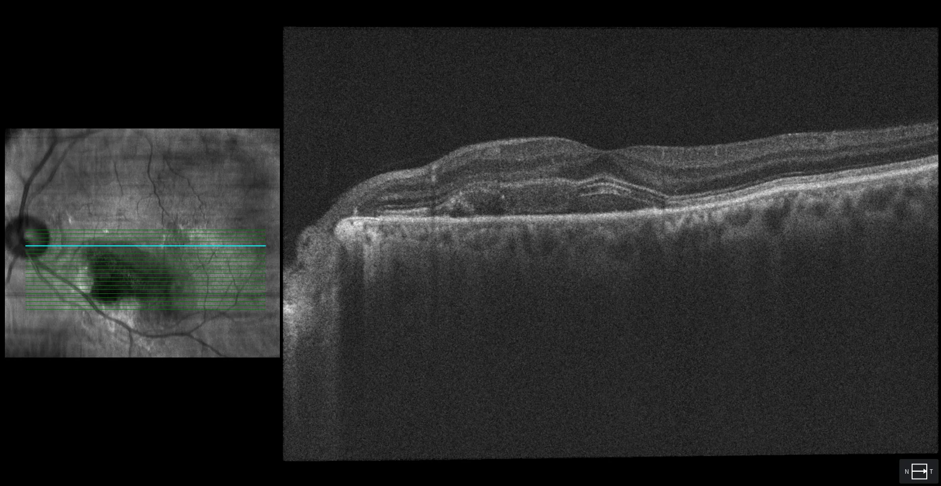
Subretinal hyperreflective material (SRHM) at foveal level corresponding to subretinal hemorrhage
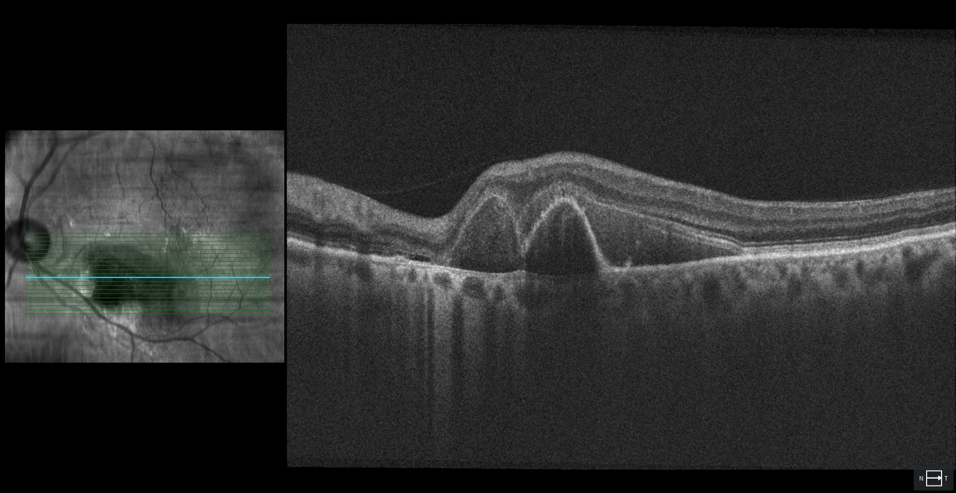
In a lower section, MHSR can also be seen, as well as 2 very pointed DEPs with hyperreflective content that suggest 2 aneurysmal dilatations with hemorrhagic content.
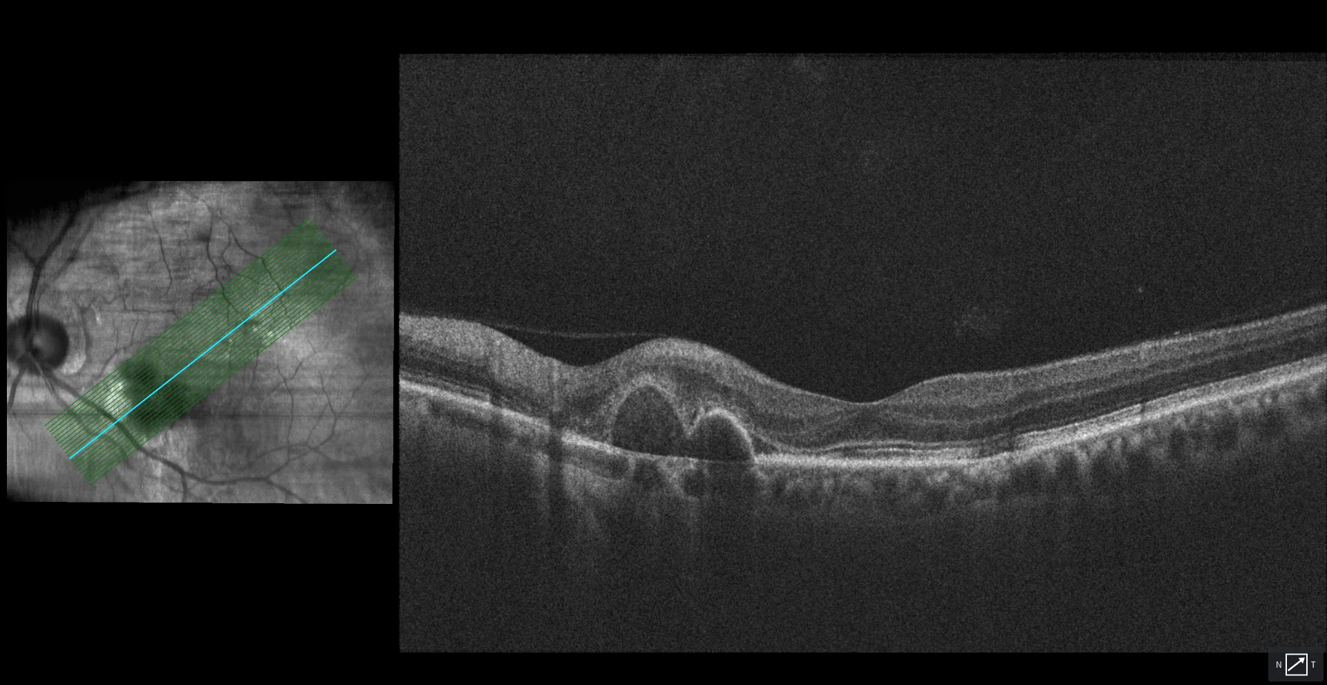
One month after the first injection with antiangiogenic, the disappearance of the subfoveal MHSR and a decrease in the height of the hemorrhagic DEPs are observed.
Description
67-year-old male who comes due to sudden loss of vision in his left eye
VA in left eye was 20/200.
The fundus showed macular hemorrhage with foveal involvement. OCT suggested the presence of aneurysmal dilatations, so antiangiogenic treatment was started. After 1 injection, VA improved to 20/20.