Macular neovascularization type 1
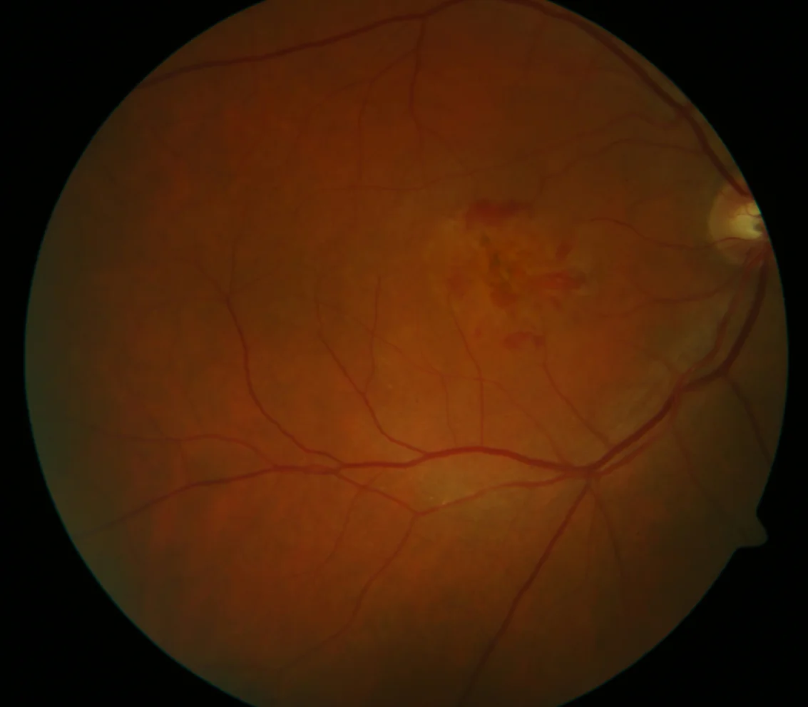
A: Yellowish macular lesion with multiple overlying hemorrhages and pigmentary changes.
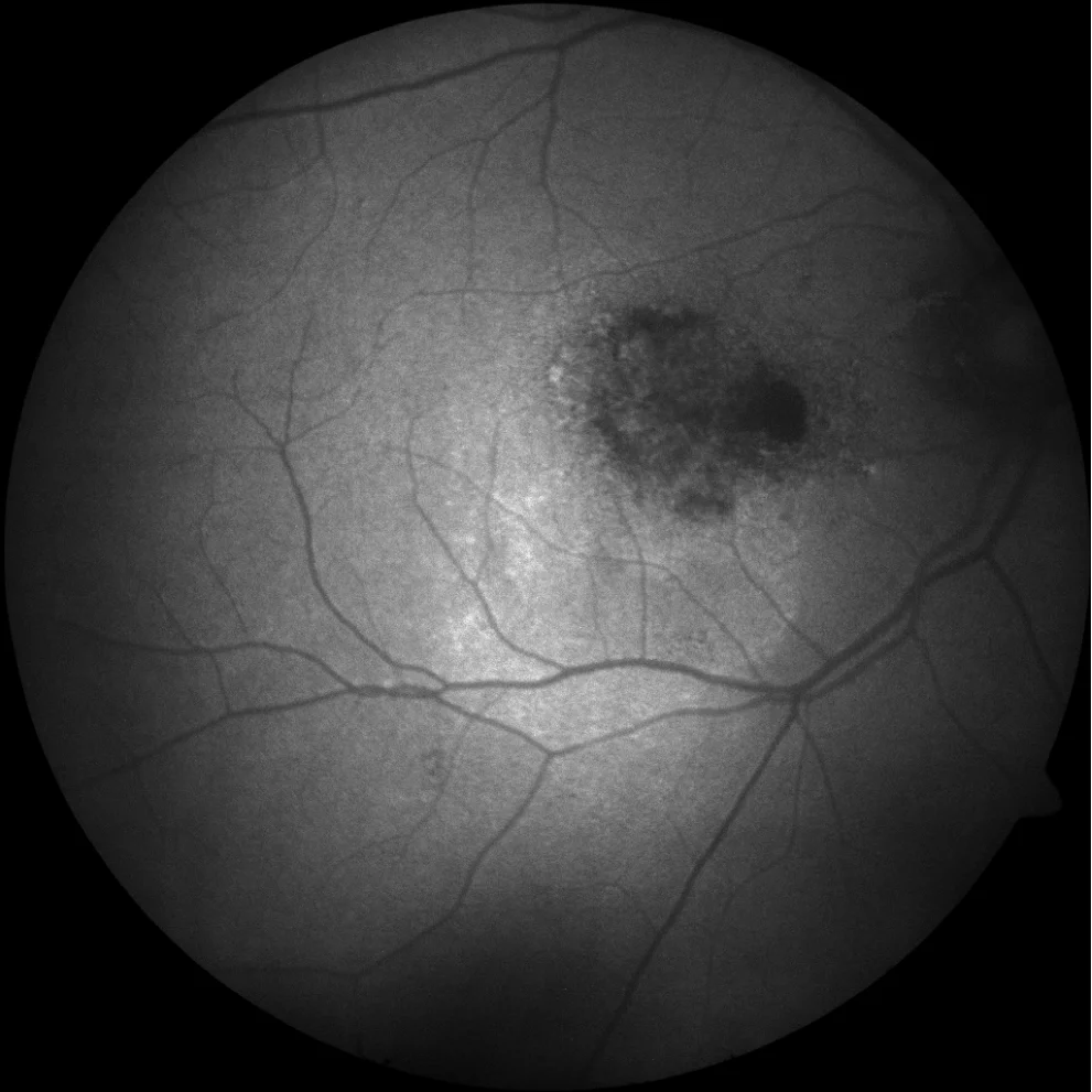
B: Blue AF showing a mottled central area (hyperAF-hypoAF) corresponding to the alteration of the RPE overlying the lesion. A small area of RPE atrophy (hypoAF) is also observed in the papillomacular bundle and a larger area of hyperAF towards the inferior temporal region corresponding to atrophy of the outer retina (unmasking effect).
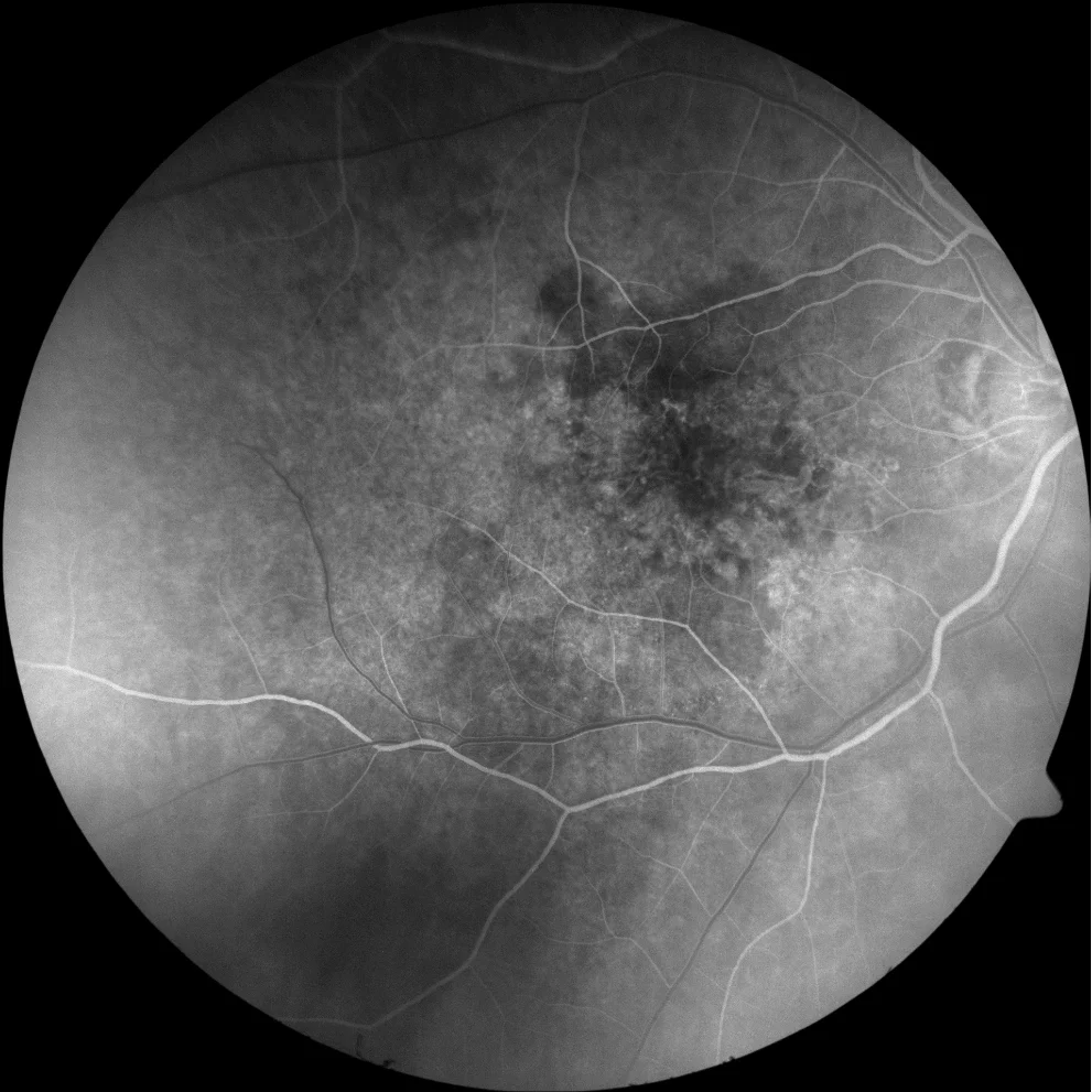
In early times a minimal mottled hyperfluorescence is observed
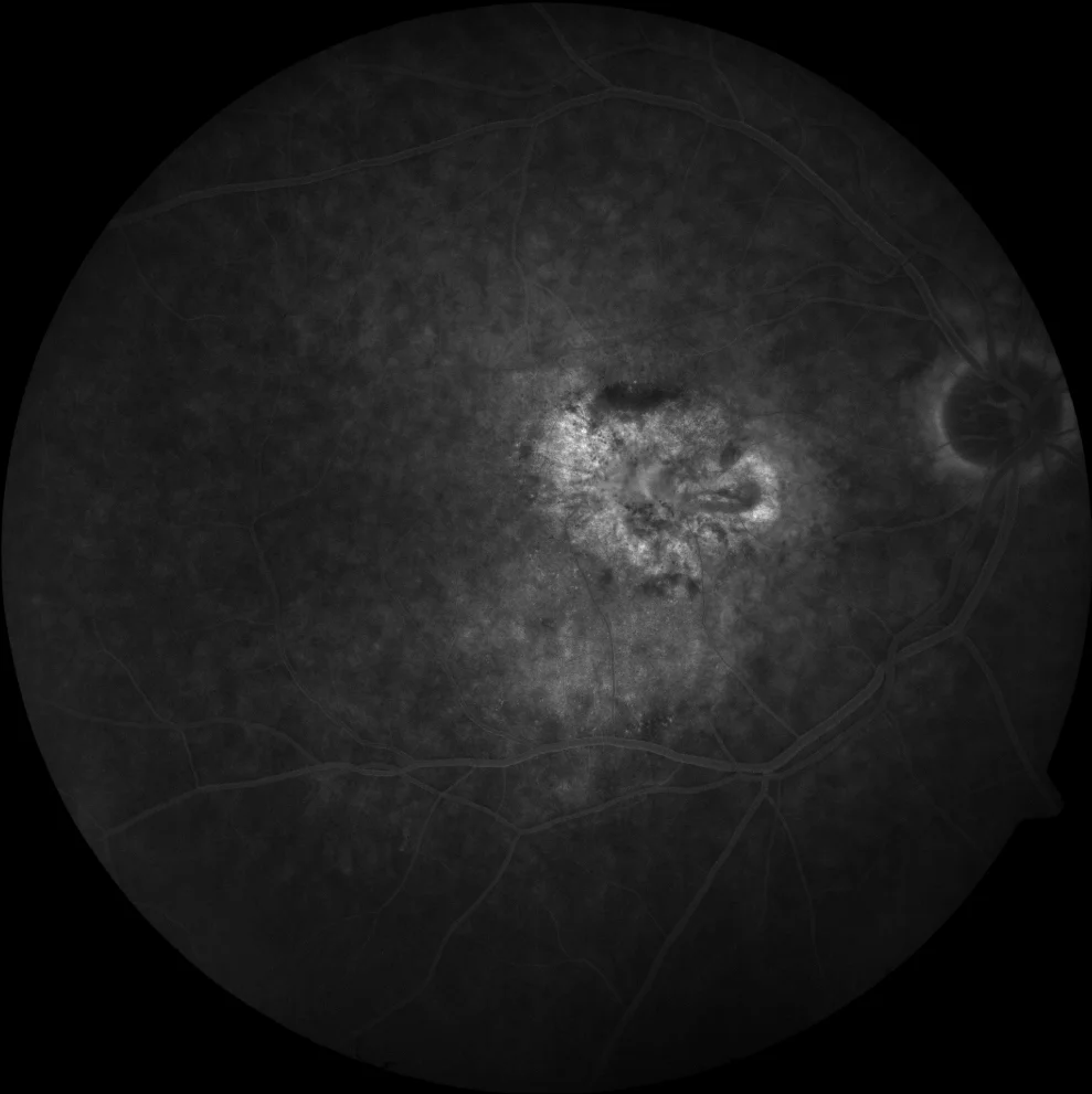
which increases in late times, but without a clear delimitation of the lesion
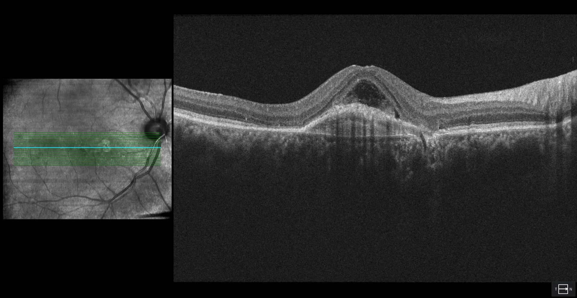
DEP with hyperreflective content corresponding to a type 1 NVM accompanied by subretinal fluid (which could even be a bacillary detachment) subfoveal. It can be seen how the nutrient vessel of the lesion crosses Bruch's membrane from the choroid in the area of RPE atrophy.
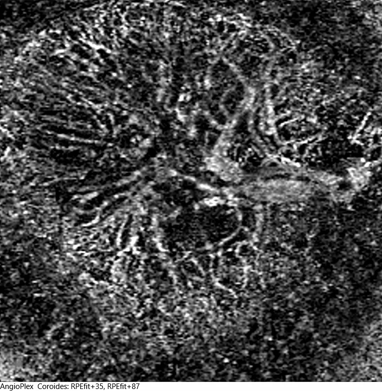
E1: Angio-OCT en face image segmenting in choroid. The neovascular lesion has many branches that emerge from a nutrient vessel at hour 3 (fan-shaped).
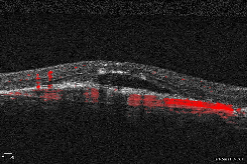
E2: In the cross section, the path below the RPE of the nutrient vessel can be seen from when it leaves the choroid until it begins to branch.
Description
73-year-old male who comes due to loss of vision in his right eye in the last month.
The BCVA in the right eye is 20/200 and in the left eye 20/25.
The fundus of the eye of the OD shows a macular lesion with multiple hemorrhages. In this case, OCT and, especially, angio-OCT, allow the diagnosis of type 1 macular neovascularization.