Macular neovascularization type 2
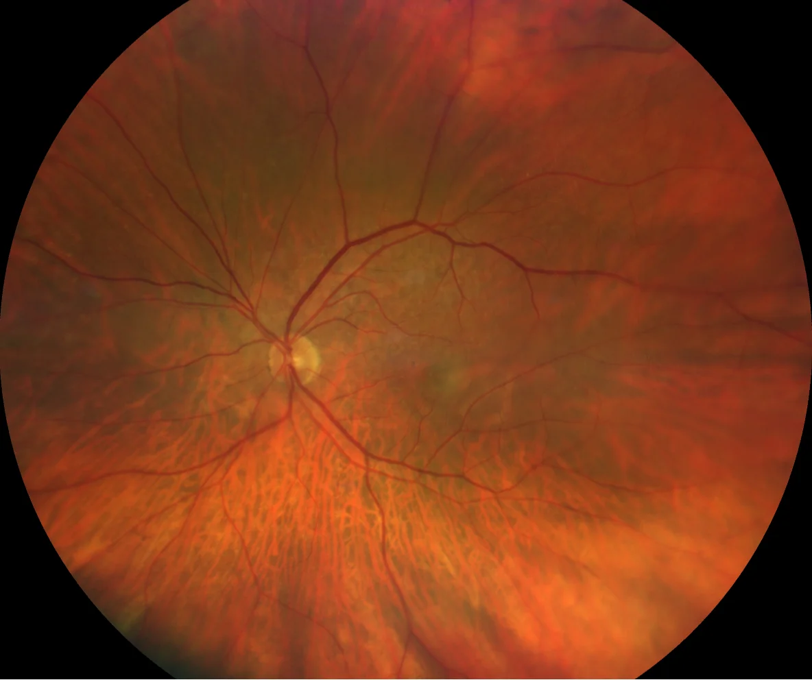
A: ill-defined greenish-yellow lesion with foveal involvement. No macular hemorrhages are observed, but reticular pseudodrusen are present, predominantly in the upper hemimacula.
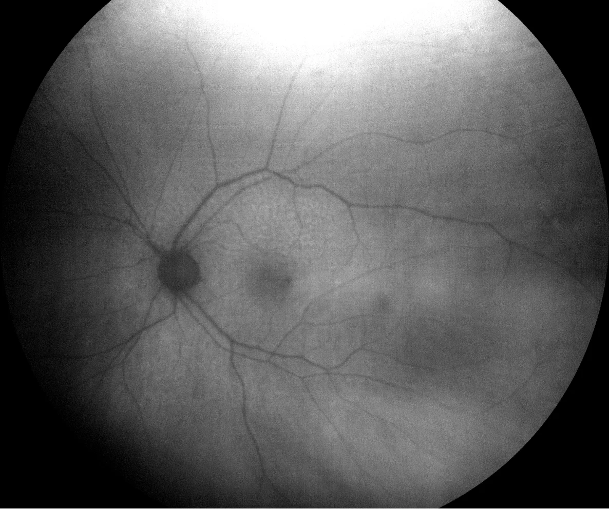
o B: Green AF showing no signal in the neovascular lesion in the LE (B1) but the characteristic punctate hypoAF (due to the screen effect) of the subretinal drusenoid deposits (SDD) in both the LE (B1) and the RE (B2).
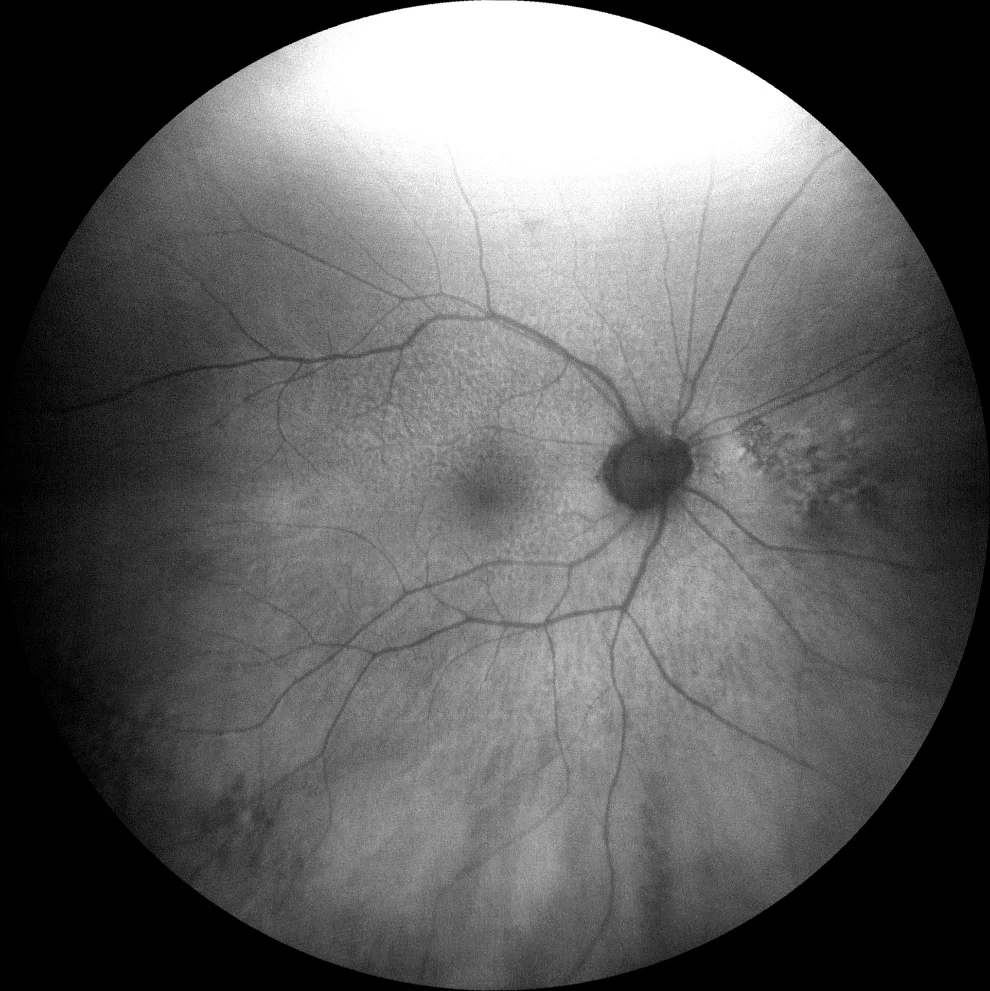
B: Green AF showing no signal in the neovascular lesion in the LE (B1) but the characteristic punctate hypoAF (due to the screen effect) of subretinal drusenoid deposits (SDD) in both the LE (B1) and the RE (B2).
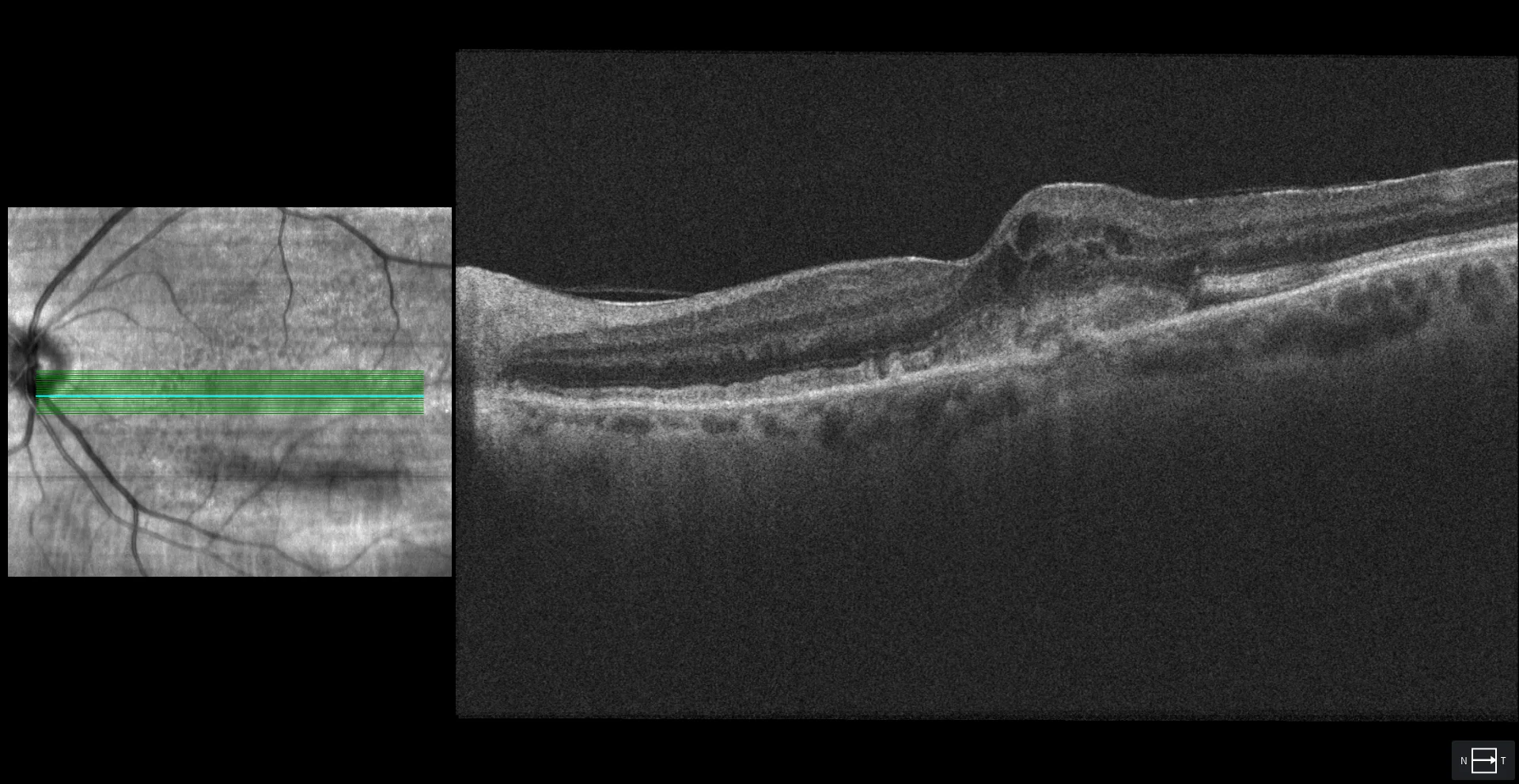
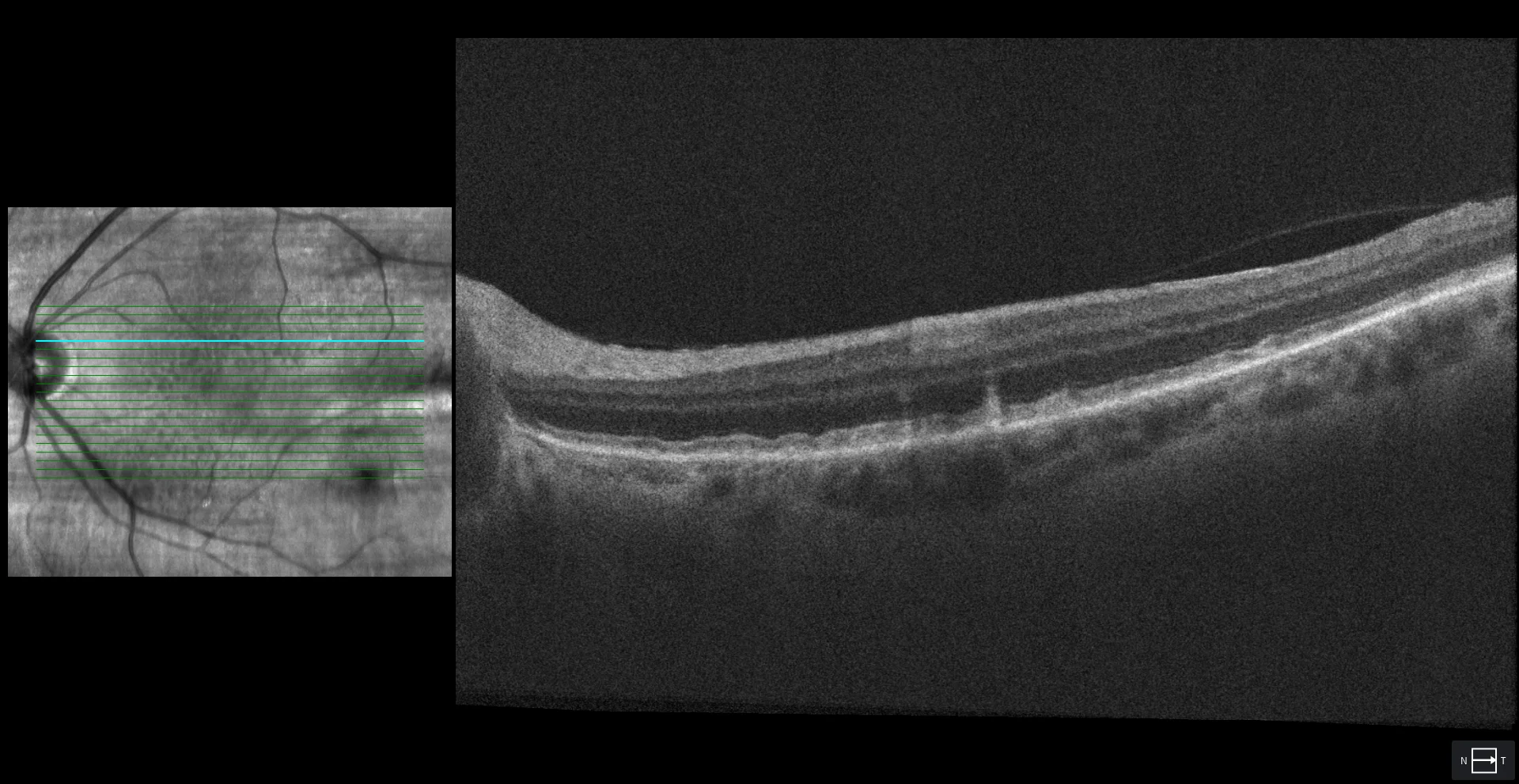
C1: Foveal section of the LE showing the subretinal hyperreflective material (SRHM) corresponding to type 2 NVM. Also observed is the break in the RPE through which the neovascular tissue penetrates from the choroid, as well as intraretinal fluid cysts (more characteristic of type 2 NVM than subretinal fluid).
SDD in both OI (C2) and OD (C3)
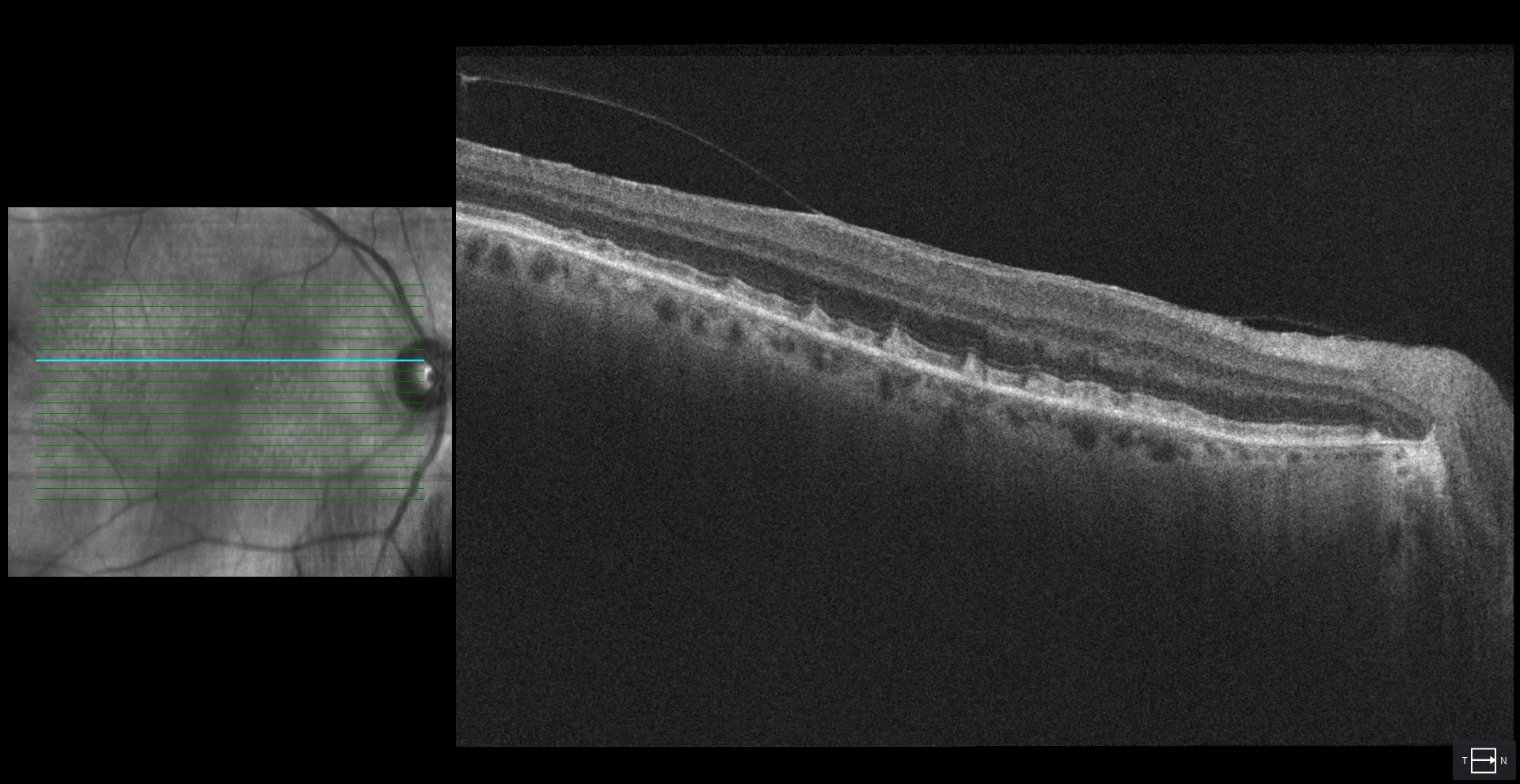
SDD in both OI (C2) and OD (C3)
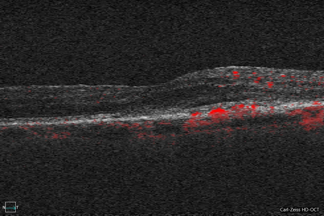
In the OCT-A cross section, D1 shows how a flow signal is detected inside the MHSR (D1). With all the cross sections and segmenting the images into the external retina and choriocapillaris, an en face reconstruction of the neovascular lesion is made (D2).
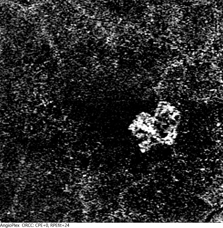
D2: In the OCT-A cross section, it can be seen that a flow signal is detected inside the MHSR (D1). With all the cross sections and segmenting the images into the external retina and choriocapillaris, an en face reconstruction of the neovascular lesion is made (D2).
Description
85-year-old woman who comes due to loss of vision in her left eye.
VA in RE is 20/25 and in LE 20/50. He has 2+ nuclear cataracts in both eyes.
The patient presented neovascular AMD with type 2 NVM, so antiangiogenic treatment was started.