Macular telangiectasia type 2
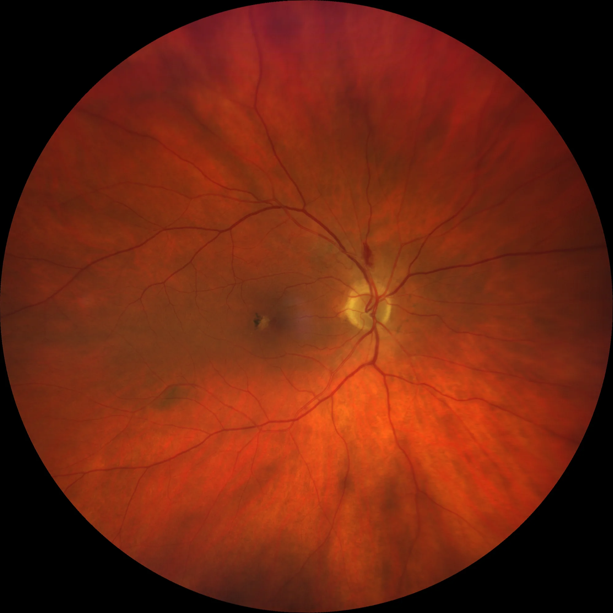
Color retinography (Clarus500, Zeiss): (A) and (B) AO: Pigmentary changes with intraretinal pigment in the macular area. In the RE, a splinter hemorrhage superior to the optic disc not related to MacTel 2 is observed.
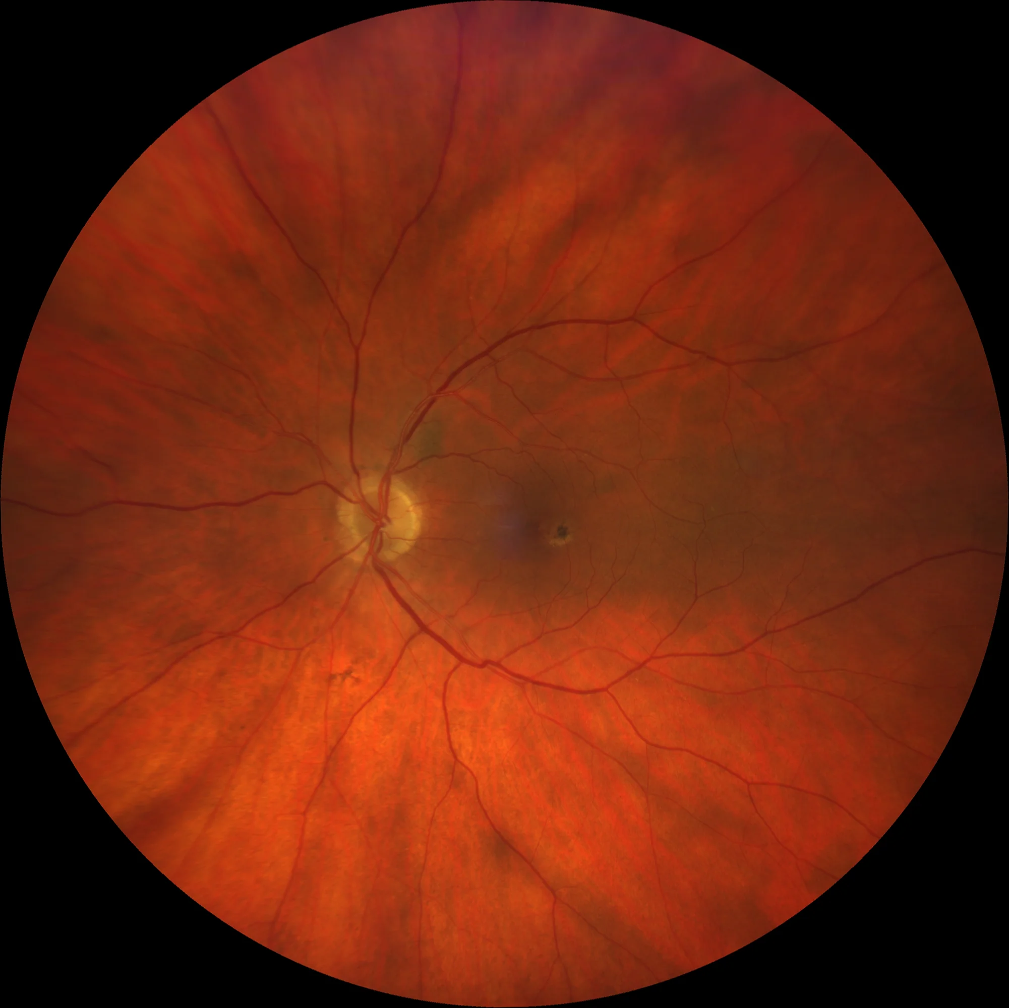
Color retinography (Clarus500, Zeiss): (A) and (B) AO: Pigmentary changes with intraretinal pigment in the macular area. In the RE, a splinter hemorrhage superior to the optic disc not related to MacTel 2 is observed.
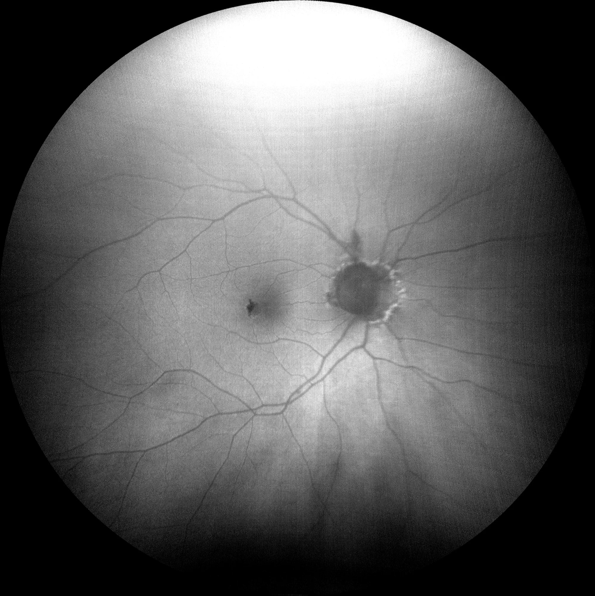
Autofluorescence (Clarus500, Zeiss): (C) and (D) AO: Hypoautofluorescence in the macular area that corresponds to the screen effect generated by RPE hyperplasia and intraretinal pigment migration.
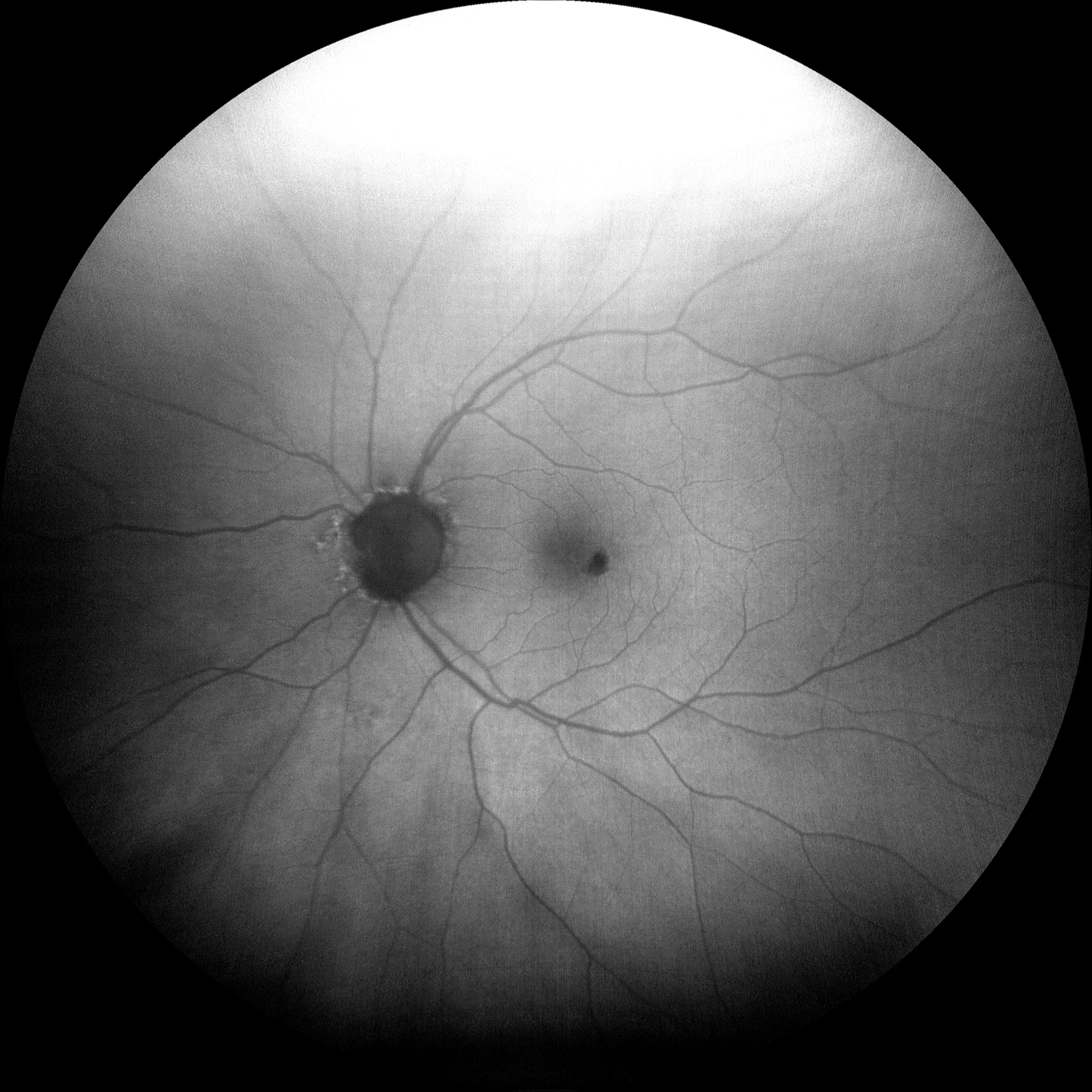
Autofluorescence (Clarus500, Zeiss): (C) and (D) AO: Hypoautofluorescence in the macular area that corresponds to the screen effect generated by RPE hyperplasia and intraretinal pigment migration.
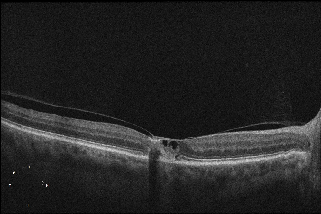
(E) Macular OCT OD: Hyporeflective cavities can be observed in the inner layers of the retina, which often correspond to degenerative areas rather than to cysts with fluid content. These cavities are called cavitations and can be present in the outer or inner retina, and sometimes only the inner boundary can be observed resting on the cavitations. Disruption of the outer layers with the presence of intraretinal hyperreflective material related to RPE hyperplasia and migration of intraretinal pigment is also observed. This patient may have developed a neovascular membrane, which is currently in a more atrophic phase. Finally, there is a vitreous-macular adhesion.
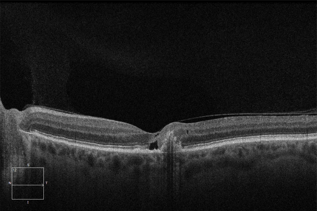
.(F) Macular OCT LE: In the LE we can also observe hyporeflective cavities but in this case in internal layers. The presence of pigmented epithelial detachment (PED) with defects in the RPE is also evident.
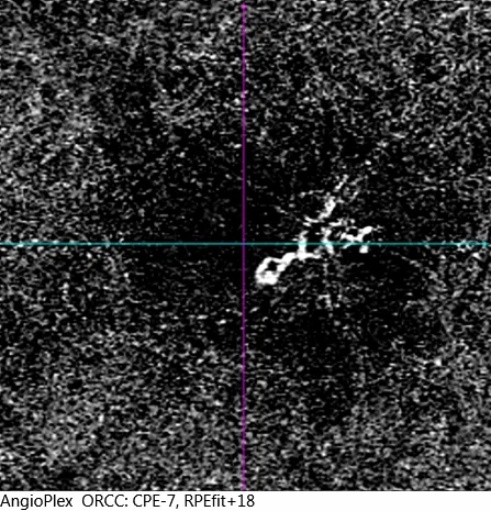
(G) Angio-OCT OI: Angio-OCT reveals the existence of a neovascular membrane in the area of the PED.
Description
Macular telangiectasia type 2. Macular telangiectasia type 2 (MacTel 2) is a type of acquired macular telangiectasia and is usually bilateral. It is the most common of the three types. Its pathophysiology involves neurodegenerative processes, vascular inflammation and vascular occlusion and ectasia phenomena that lead to degeneration and loss of outer retinal layers that can end up forming lamellar pseudoholes and formation of cystic-looking cavities, telangiectatic vessels in the macular area, hyperplasia of the retinal pigment epithelium (RPE) and migration to internal layers, appearance of crystalline deposits below the internal limiting membrane and development of neovascular membranes in advanced stages.
The following classification has been proposed:
- Stage 1: Normal fundoscopic examination. Very subtle changes in autofluorescence.
- Stage 2: Loss of retinal transparency. Hyperautofluorescence. Mild telangiectatic changes without exudation. Sublimiting internal crystalline deposits.
- Stage 3: Moderate telangiectatic changes. Right-angled venules and capillary remodeling in the outer layers of the retina, without exudation.
- Stage 4: Pigmentary changes due to hyperplasia and intraretinal migration of RPE cells. Degenerative changes.
- Stage 5: Reino-choroidal anastomosis with exudation or hemorrhages.