Macular telangiectasias type 2 (MacTel2)
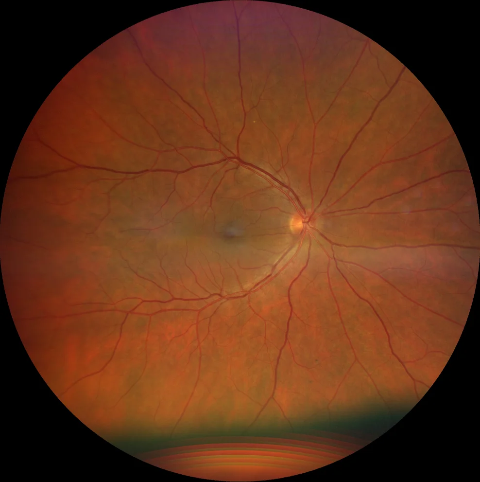
Retinography (Clarus 700, Zeiss): macular appearance practically normal in both eyes (A1, A2). No right-angled vessels are observed.
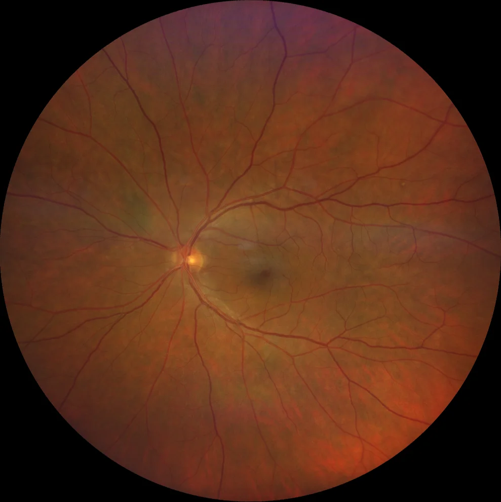
Retinography (Clarus 700, Zeiss): macular appearance practically normal in both eyes (A1, A2). No right-angled vessels are observed.
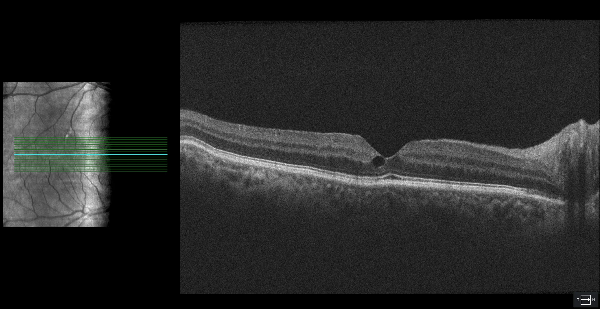
OCT (Cirrus 5000, Zeiss): disorganization of the internal retinal layers and pseudocyst in the area temporal to the fovea in both eyes (B1, B2). In the RE there is a discontinuous MLE in this area (B1). In the LE not only the MLE is discontinuous but also the EZ, which justifies the patient's poorer vision in this eye (B2).
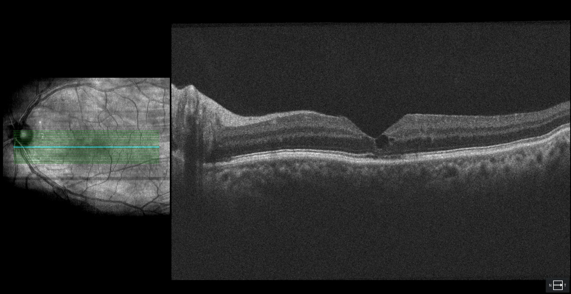
OCT (Cirrus 5000, Zeiss): disorganization of the internal retinal layers and pseudocyst in the area temporal to the fovea in both eyes (B1, B2). In the RE there is a discontinuous MLE in this area (B1). In the LE not only the MLE is discontinuous but also the EZ, which justifies the patient's poorer vision in this eye (B2).
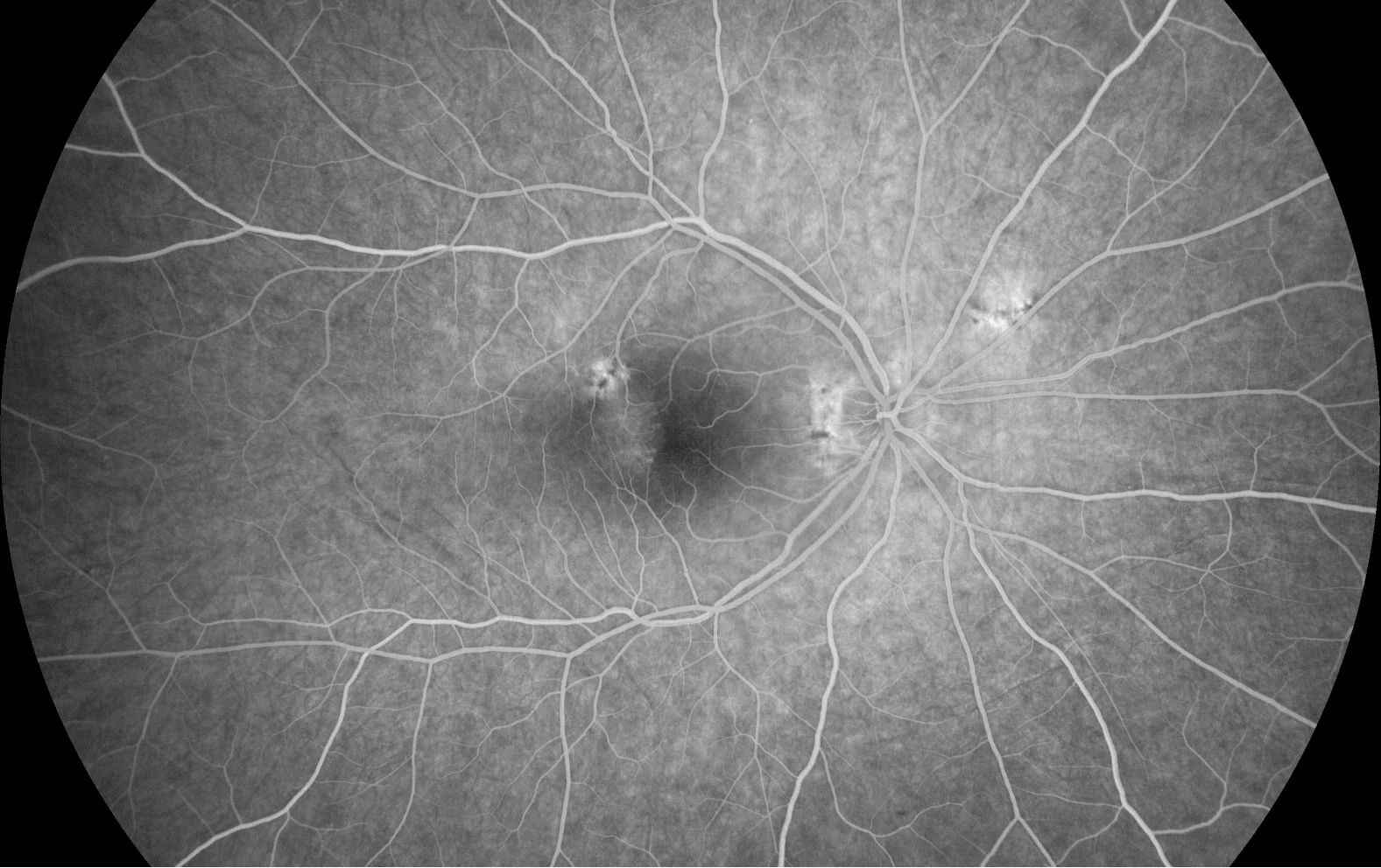
AGF (Clarus 700, Zeiss): lesions with early hyperfluorescence (C1, C2) and late contrast leakage (C3, C4), which is suggestive of vascular injury. The symmetry of the lesions confirms the diagnosis of MacTel2.
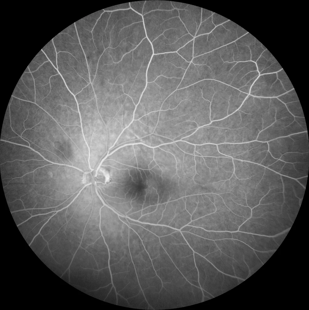
AGF (Clarus 700, Zeiss): lesions with early hyperfluorescence (C1, C2) and late contrast leakage (C3, C4), which is suggestive of vascular injury. The symmetry of the lesions confirms the diagnosis of MacTel2.
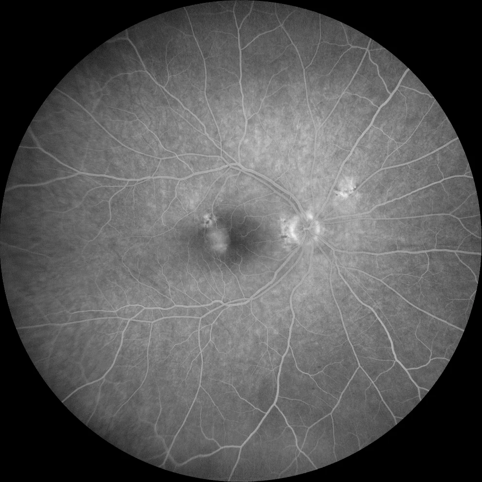
AGF (Clarus 700, Zeiss): lesions with early hyperfluorescence (C1, C2) and late contrast leakage (C3, C4), which is suggestive of vascular injury. The symmetry of the lesions confirms the diagnosis of MacTel2.
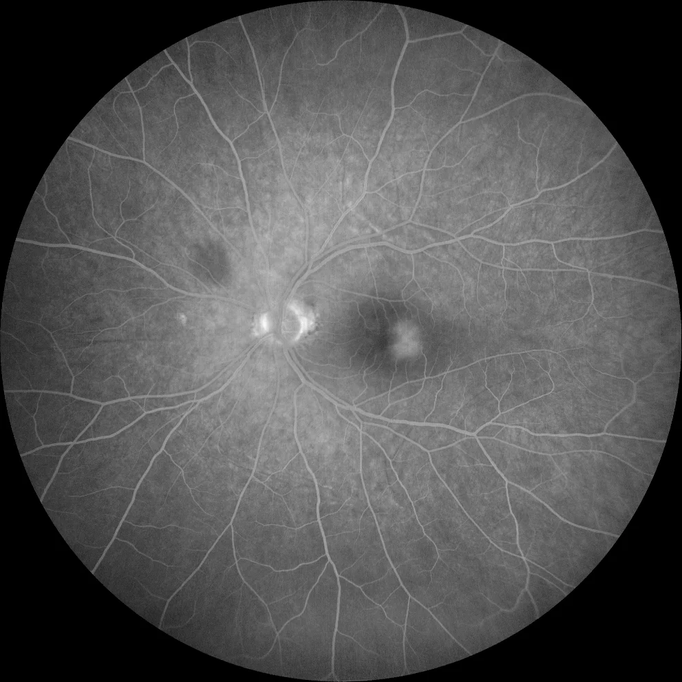
AGF (Clarus 700, Zeiss): lesions with early hyperfluorescence (C1, C2) and late contrast leakage (C3, C4), which is suggestive of vascular injury. The symmetry of the lesions confirms the diagnosis of MacTel2.
Description
A 65-year-old male with a history of type 2 diabetes mellitus on metformin treatment presents with progressive vision loss in both eyes.
Visual acuity is 20/25 in the right eye and 20/30 in the left eye. The fundus has a practically normal appearance. OCT of the right eye shows disorganization of the internal retinal layers, a pseudocyst and a discontinuous external limiting membrane (ELM) in the area temporal to the fovea. OCT of the left eye shows the same disorganization of the internal layers, a pseudocyst, as well as discontinuity in the ELM and the ellipsoid zone (EZ), also in the area temporal to the fovea. Fluorescein angiography shows early hyperfluorescence with contrast leakage at late times in these areas temporal to the fovea. A diagnosis of idiopathic macular telangiectasias type 2 (MacTel2) is made.