Myopia Case 1 Tiled Background
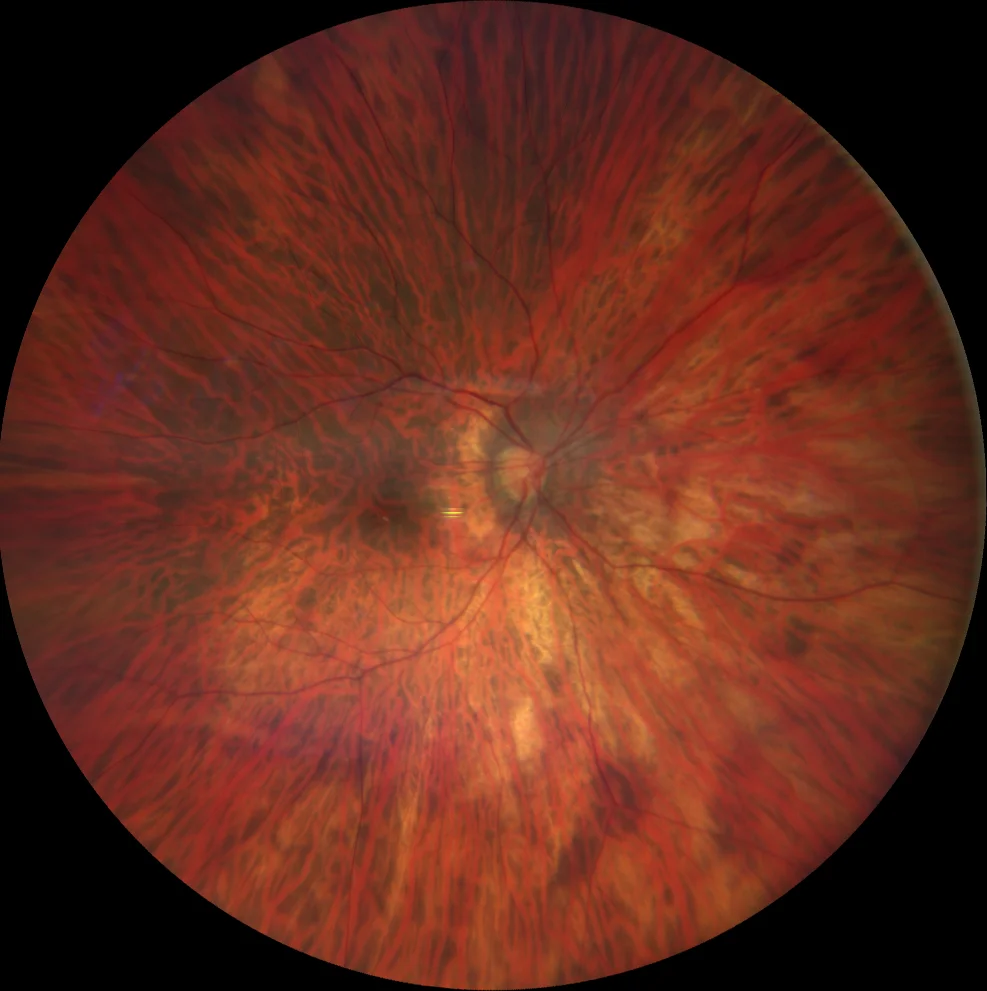
Retinography (Clarus 700, Zeiss): visualization of the large choroidal vessels in the macula and in the periphery 360º in both eyes
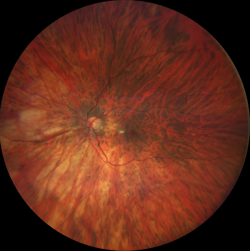
Retinography (Clarus 700, Zeiss): visualization of the large choroidal vessels in the macula and in the periphery 360º in both eyes
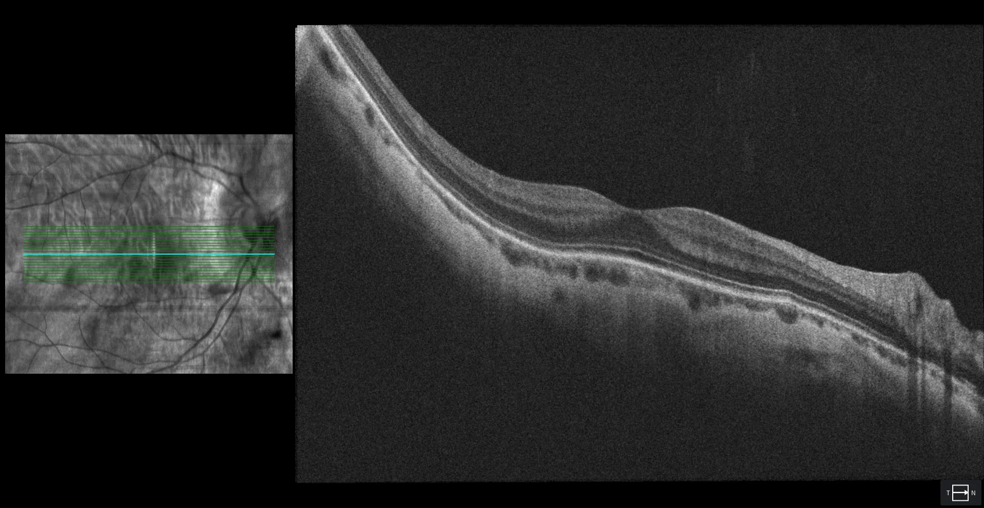
OCT (Cirrus 5000-HD, Zeiss): significant thinning of the stromal choroid in both eyes. No tractional or neovascular component is observed in the context of the patient's pathological myopia.
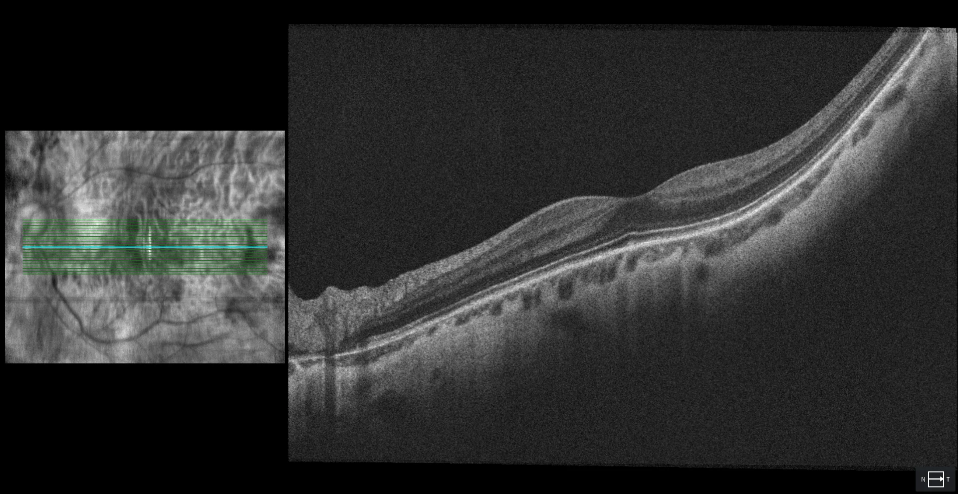
OCT (Cirrus 5000-HD, Zeiss): significant thinning of the stromal choroid in both eyes. No tractional or neovascular component is observed in the context of the patient's pathological myopia.
Description
43-year-old woman who attends regular check-ups.
AV OD 20/25 with -18.50.
AV OI 20/25 with -19.00.
A very tessellated fundus is observed in both eyes, as well as a small peripapillary staphyloma in the right eye. No other macular or peripheral alterations are observed, either in the retinography or in the OCT.