Myopic retinoschisis
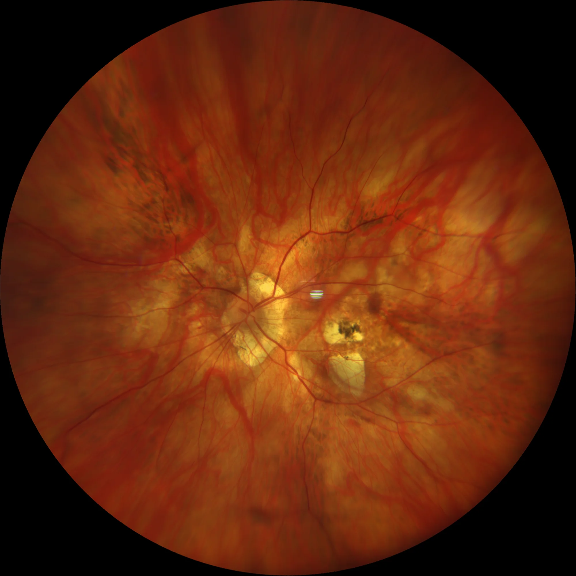
LE: High myopic fundus with two small juxtamacular plaques of chorioretinal atrophy. A pigmented lesion corresponding to a Fuchs spot is observed on the plaque of atrophy closest to the macula.
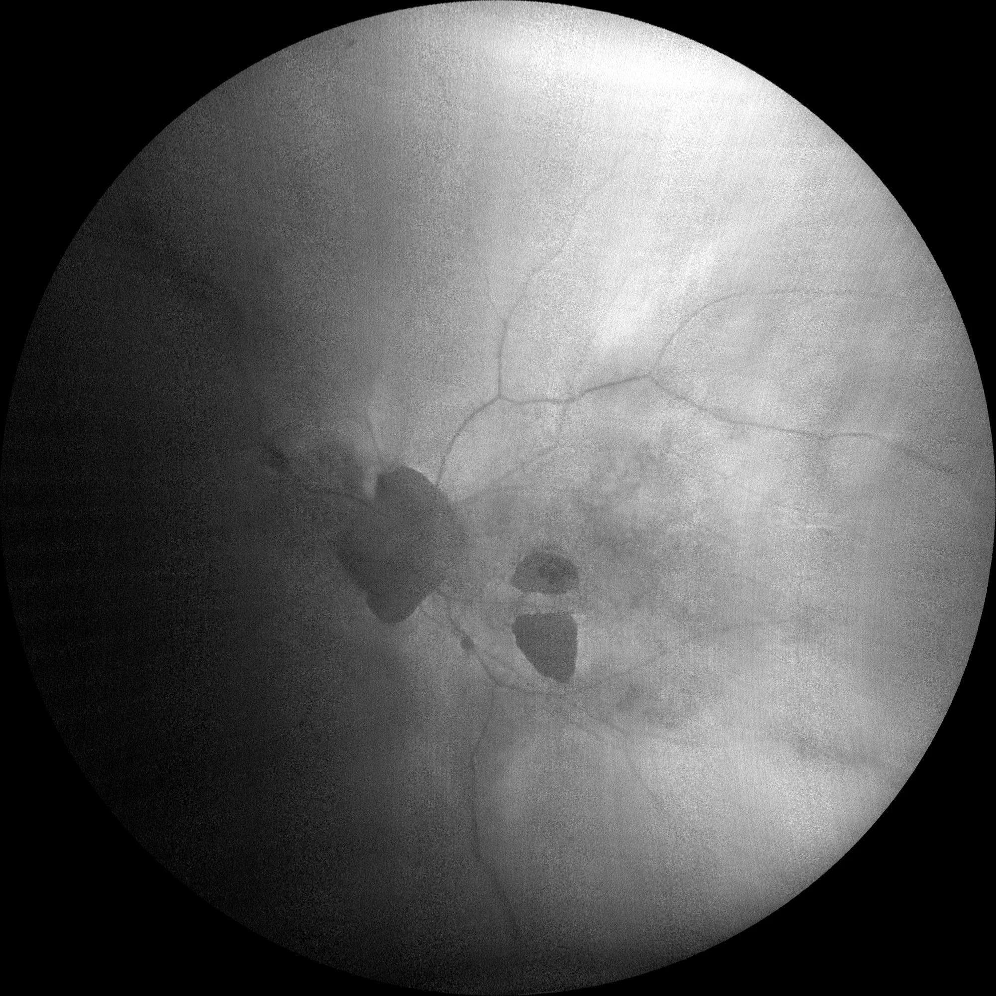
Two hypoautofluorescent plaques are observed in the posterior pole and another peripapillary plaque that correspond to the chorioretinal atrophy plaques.
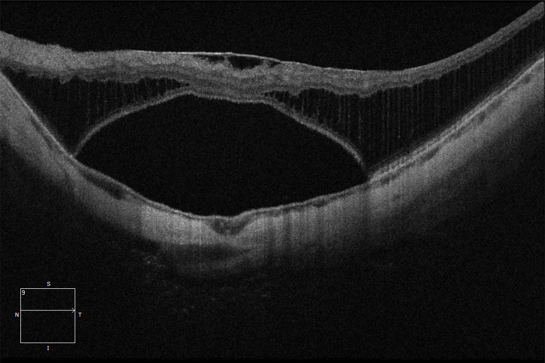
OI: Tractional myopic maculopathy with the presence of retinoschisis, foveal detachment and epiretinal membrane. It corresponds to stage T3 of the ATN classification of tractional myopic maculopathy.
Description
Progressive separation of the retinal layers caused by the existence of a posterior staphyloma that generates an external traction, in combination with the presence of internal tractions caused by retinal rigidity and the internal limiting membrane, vitreous adhesions or epiretinal membranes. The ATN classification distinguishes 6 different categories within the tractional component:
- T0: Without macular retinochisis
- T1: Internal or external foveoschisis
- T2: Internal + external foveoschisis
- T3: Foveal detachment
- T4: Macular hole
- T5: Macular hole + Retinal detachment