Neovascular membrane associated with optic nerve drusen
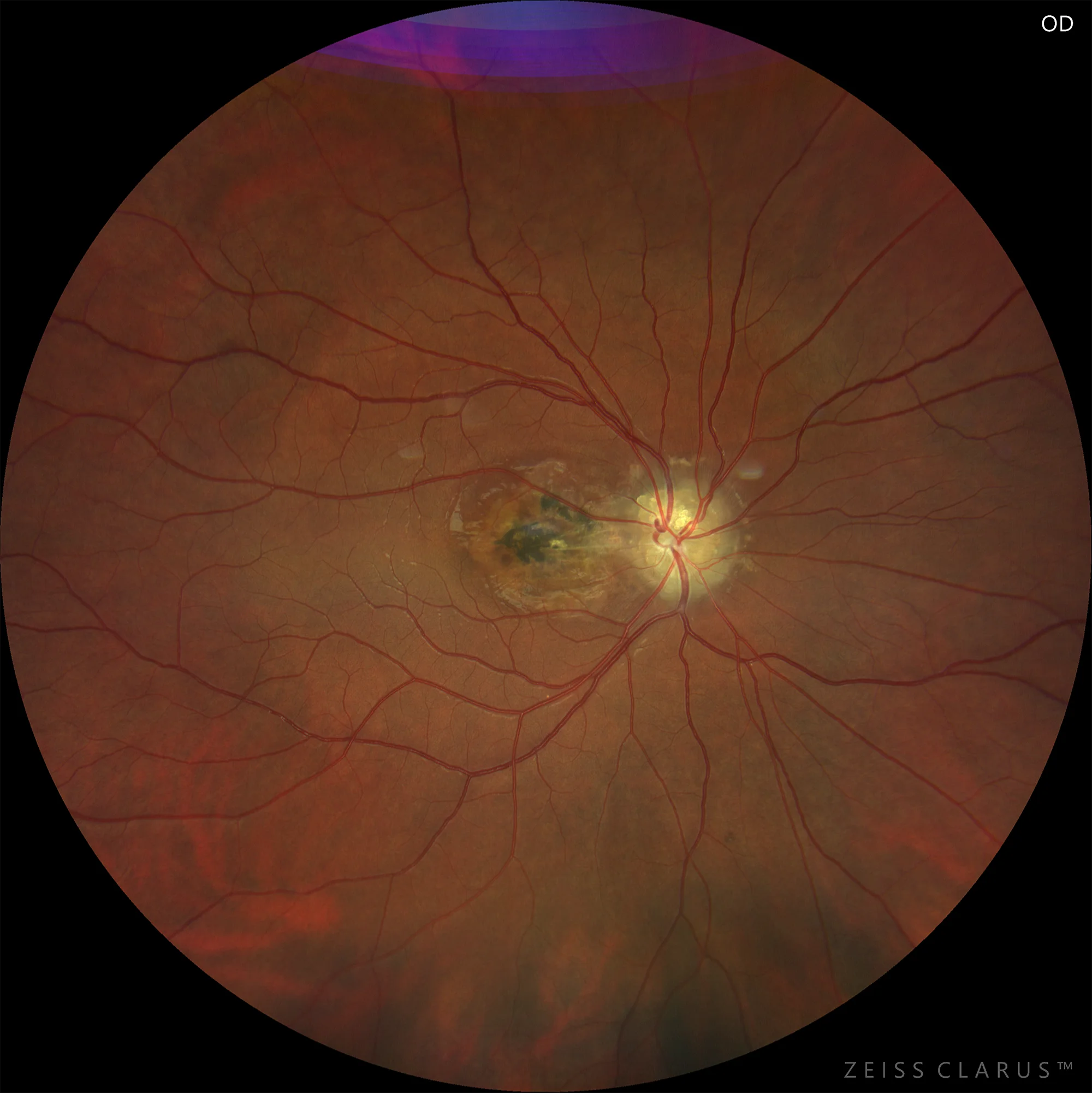
Figure 1. Retinography of the right eye. Superficial optic nerve drusen, fibrous neovascular membrane in the papillomacular bundle with pigment hypertrophy and halo of atrophy of the perilesional pigment epithelium, reaching the foveal area.
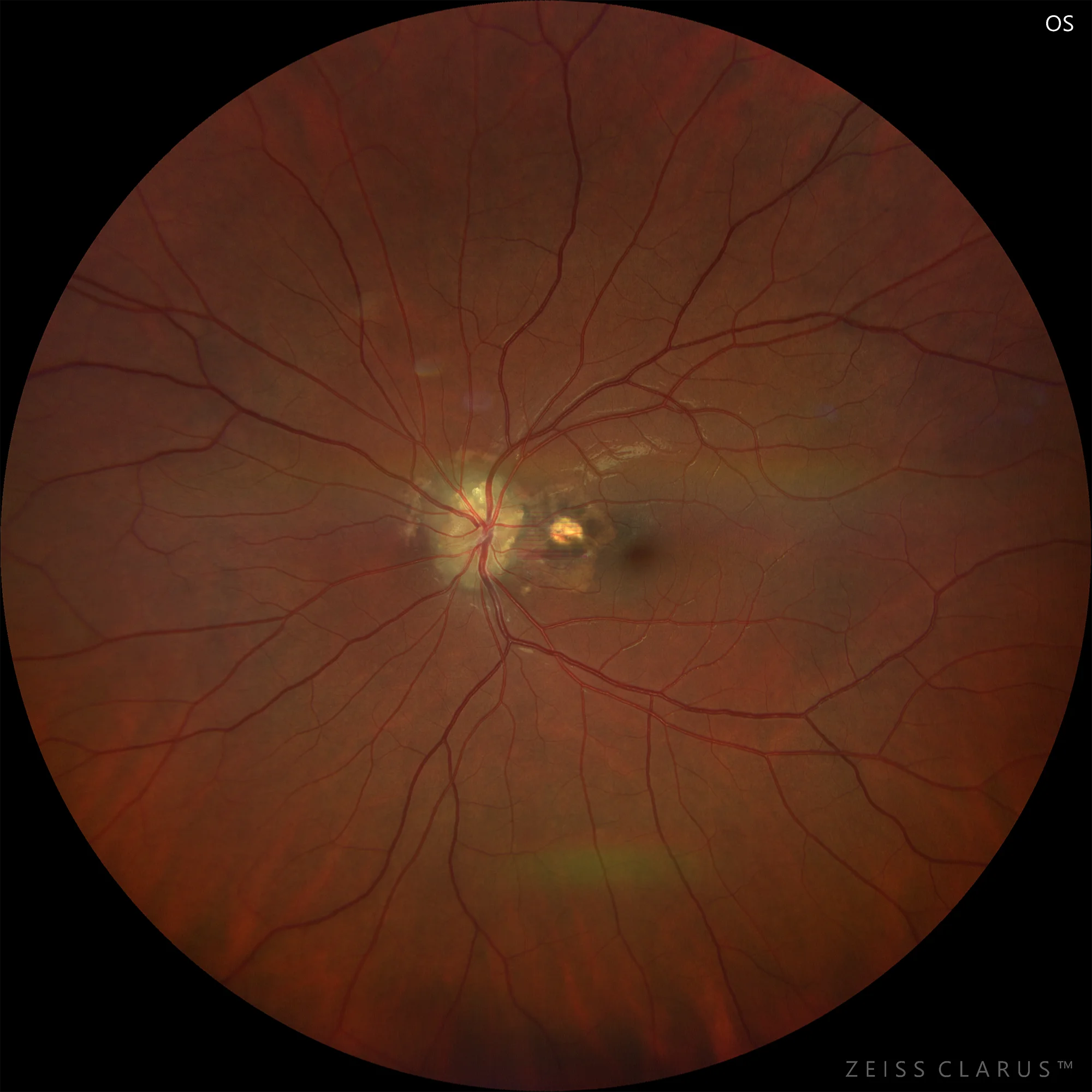
Figure 2. Retinography of the left eye. Superficial optic nerve drusen, fibrous neovascular membrane in the papillomacular bundle, with a halo of retinal pigment epithelium atrophy that does not reach the fovea.
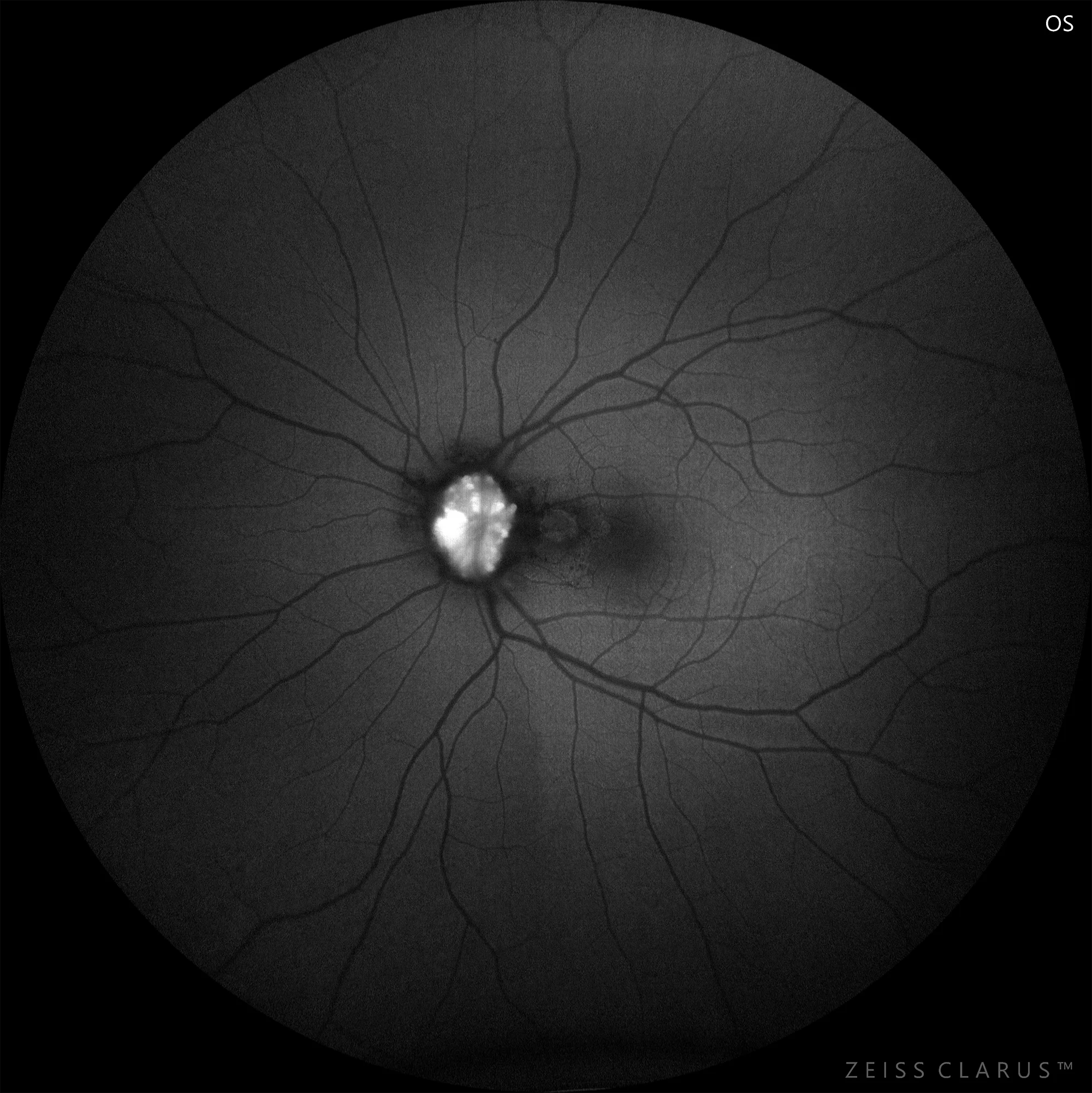
Figure 3. Autofluorescence of the left eye
Description
Characteristics:
Optic nerve drusen (OND) are rounded structures composed of calcium salts and other proteins that develop in the optic nerve and evolve dynamically in adulthood. They usually appear in small optic discs, probably due to axonal transport blockage, and are bilateral and asymmetric in 75% of cases. Diagnosis is based on biomicroscopy, and autofluorescence will confirm the diagnosis, especially in the case of buried drusen. In most cases, OND are asymptomatic, but progressive visual loss may occur secondary to papillary congestion. Rarely, vascular complications may appear, such as choroidal neovascularization, ischemic optic neuritis, occlusion of the central retinal artery or vein, or retinal or optic nerve hemorrhages.