Neovascular membrane type 3
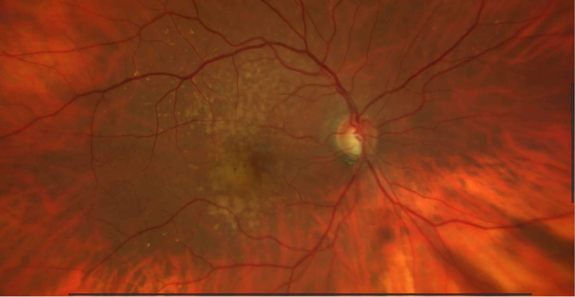
Retinography: In both eyes, soft confluent drusen are seen in the macular area. In the right eye, two intraretinal punctate hemorrhages are seen in the macular area.
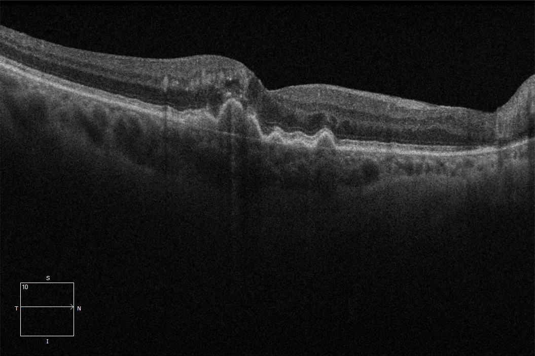
Macular OCT: Drusenoid detachments of the RPE in both eyes. In the right eye, juxtafoveal intraretinal hyperreflectivity is seen due to intraretinal neovascularization with minimal associated cystic edema.
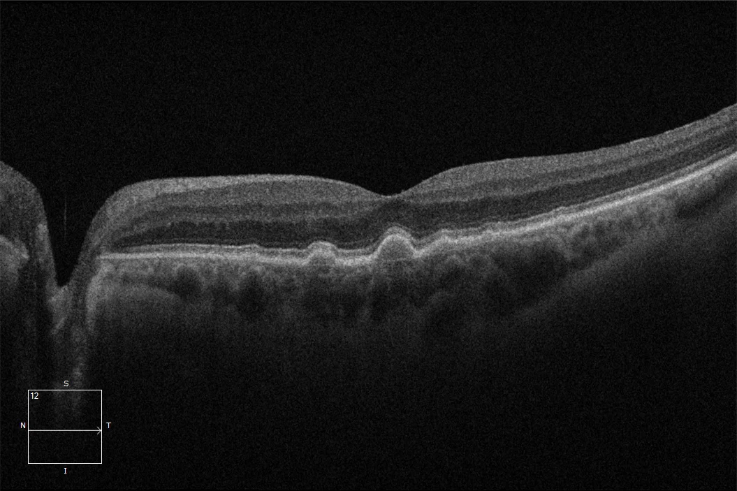
Macular OCT: Drusenoid detachments of the RPE in both eyes. In the right eye, juxtafoveal intraretinal hyperreflectivity is seen due to intraretinal neovascularization with minimal associated cystic edema.
Description
Neovascular age-related macular degeneration (AMD) accounts for approximately 1 in 10 cases of AMD. In particular, in type 3 neovascularization, neovessels originate from the capillary plexuses of the retina and secondarily invade the subretinal space, crossing the retinal pigment epithelium (RPE). Type 3 neovascularization, also known as retinal angiomatous proliferation, is classified into three stages of progression. In stage I, as in the case described, neovascular proliferation begins in the deep capillary plexus of the retina and remains limited to the macula, without affecting the submacular space.