Nonproliferative diabetic retinopathy
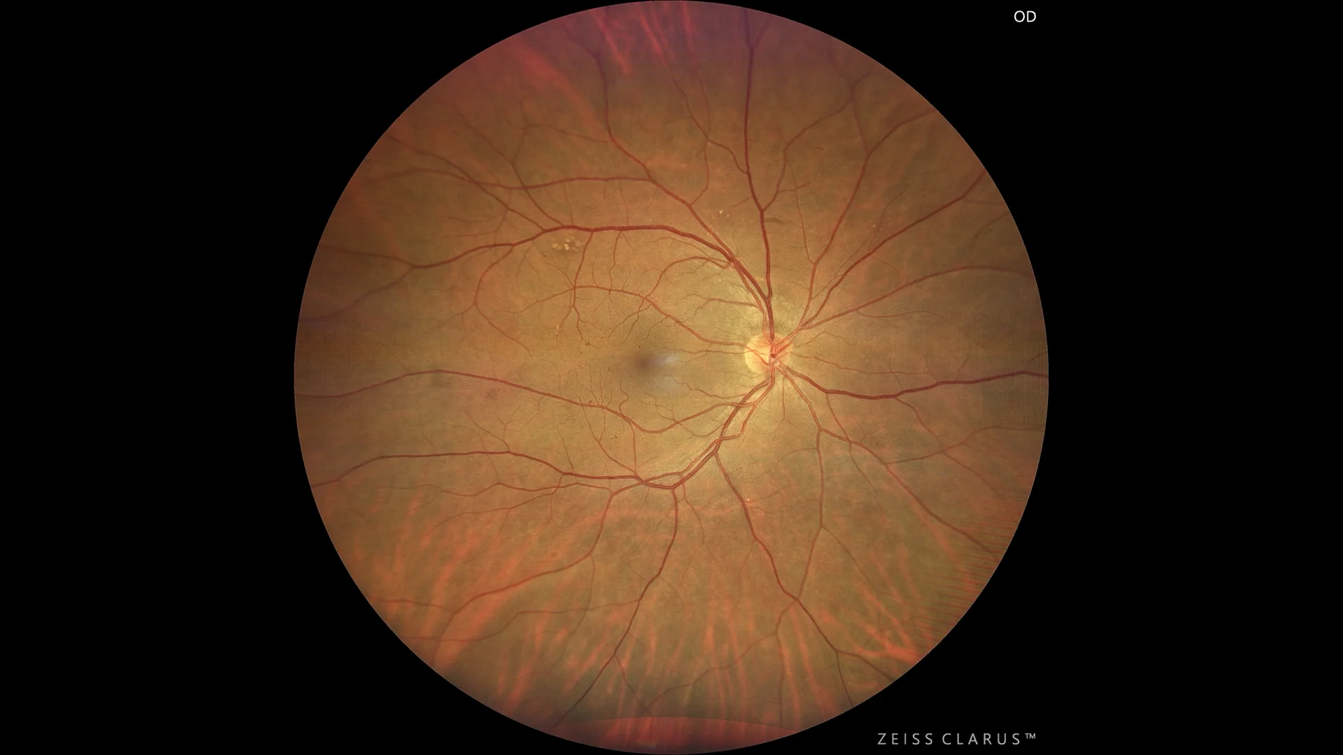
Color retinography in which some microaneurysms can be seen in the posterior pole, as well as some dispersed lipid exudates that do not threaten the macula.
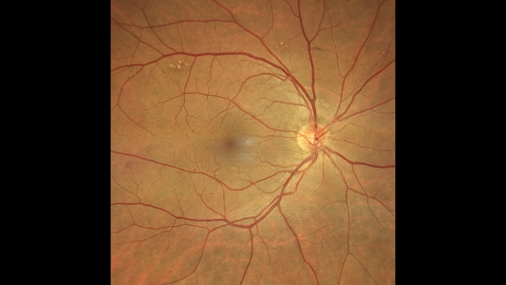
Color retinography enhanced in the posterior pole to visualize microaneurysms and other vascular changes in more detail.
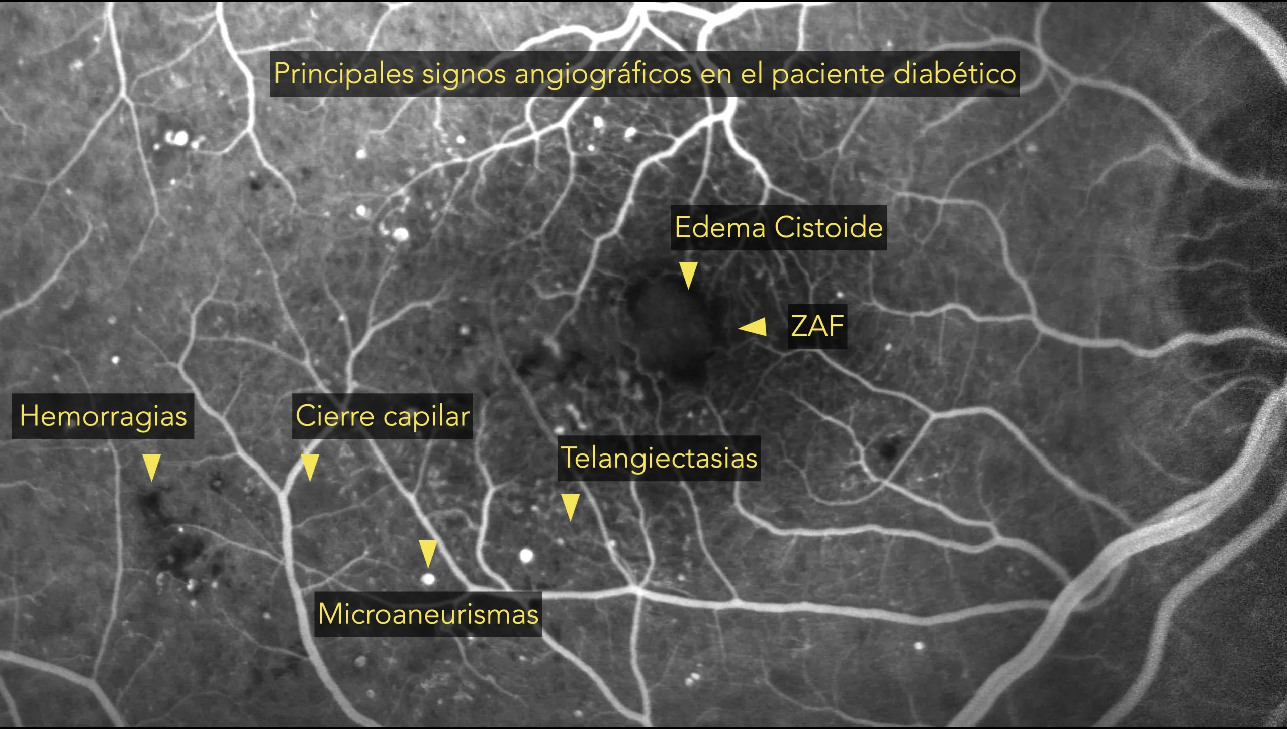
33º fluorescein angiography in which we can observe the main findings that accompany diabetic retinopathy.
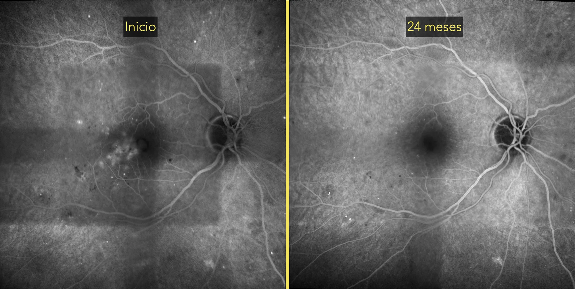
55º fluorescein angiography where we can see the before and after after 2 years of intravitreal treatment with antiangiogenics.
Description
In nonproliferative diabetic retinopathy, findings on fundusography include microaneurysms, small dilations in blood vessels; retinal hemorrhages in the form of spots or dots; hard exudates, which are accumulations of lipids and proteins; and areas of retinal thickening due to fluid buildup (macular edema). These changes are indicative of vascular damage caused by diabetes and can be monitored by regular fundus examinations to assess disease progression.