Optic nerve sheath meningioma
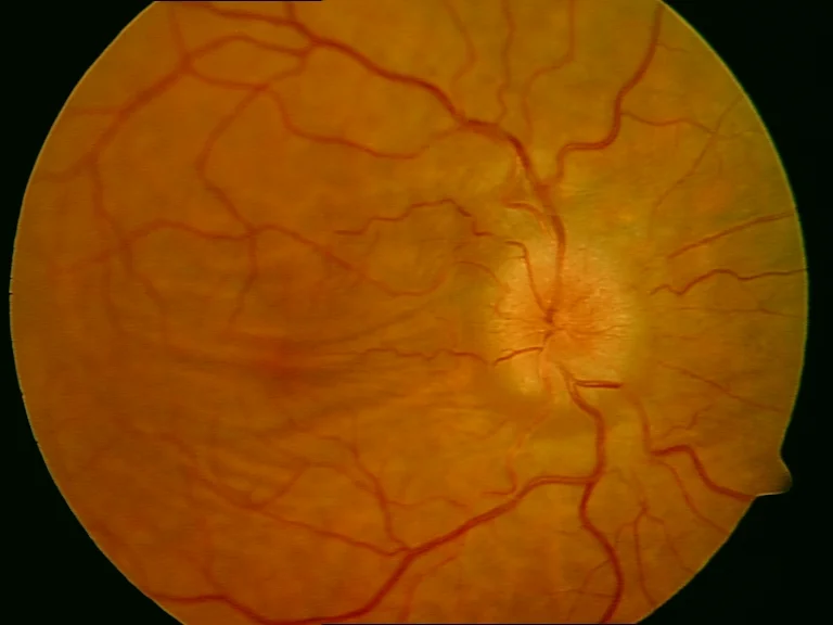
Retinography (FF450 IR plus Zeiss)
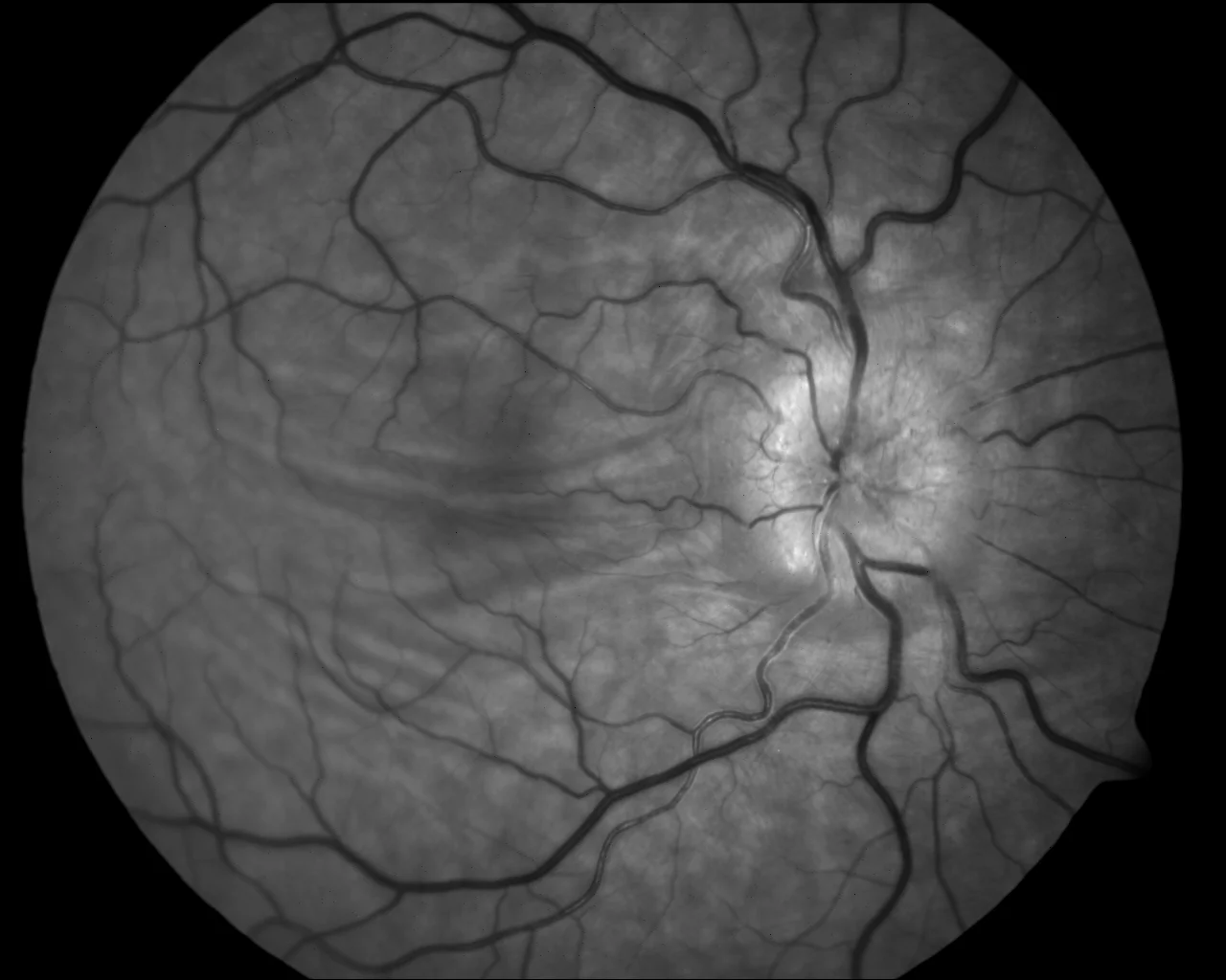
Color and green filter: Papilla edema, chorioretinal folds and vascular tortuosity
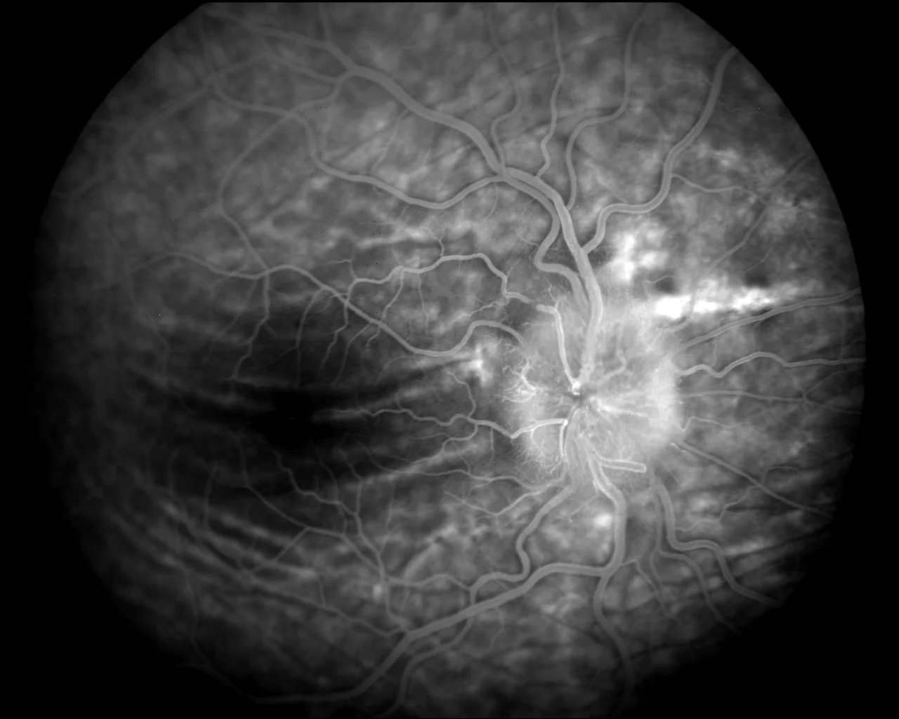
AGF: Papillary oozing (papilla edema) stands out.
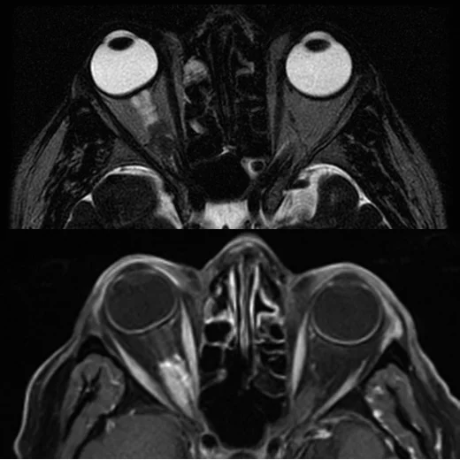
Right optic nerve sheath meningioma with diffuse tubular thickening, compression and deformity of the eyeball. The tumor enhances with intravenous contrast injection.
Description
Optic nerve sheath meningioma is a rare benign neoplasm that arises from the meningoendothelial cells surrounding the optic nerve. It is the second most common primary tumor of the optic nerve, accounting for 1-2% of all meningiomas. The incidence is highest in adult women in their 4th-5th decade. Although considered benign tumors, primary meningiomas cause slow, progressive vision loss secondary to compression of the optic nerve and its blood supply.
The diagnosis is confirmed with MRI, which allows confirming the characteristic pattern and assessing the degree of extension.