Optic nerve tumor pathology – melanocytoma
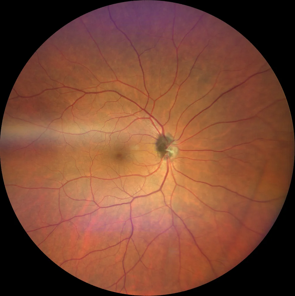
Retinography (Clarus 700, Zeiss): pigmented lesion in the optic nerve head extending to the adjacent retina superiorly (A).
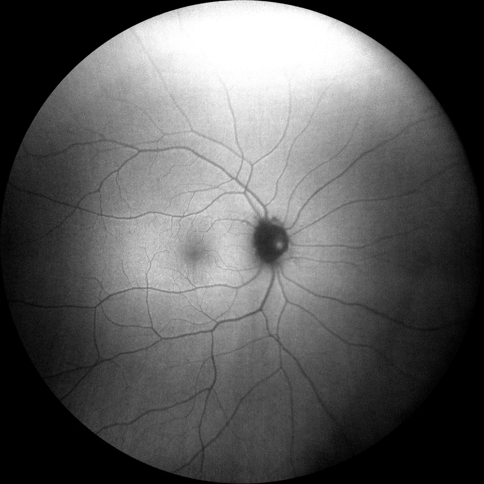
Green autofluorescence (Clarus 700, Zeiss): the lesion is hypoautofluorescent (B).
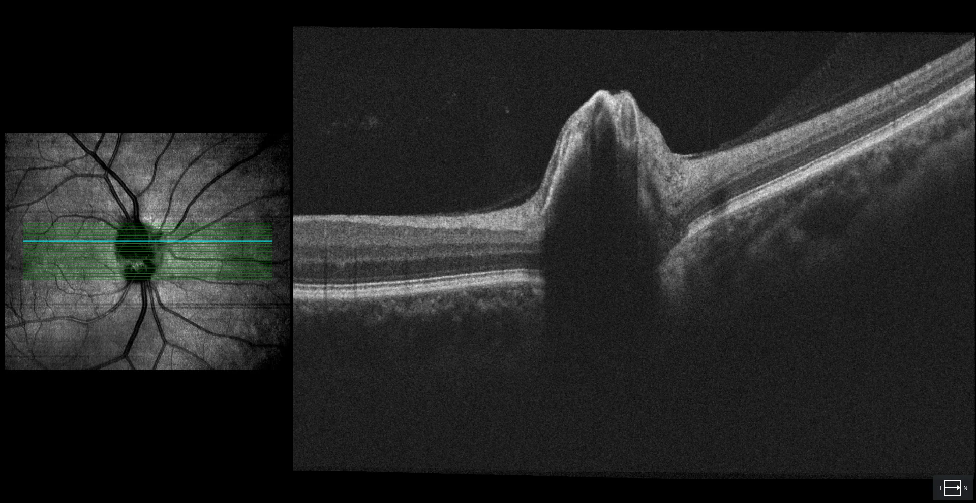
OCT (Cirrus 5000, Zeiss): superficial lesion with intense posterior shadow (C).
Description
55-year-old asymptomatic woman who comes for a check-up.
Visual acuity is 20/20 in both eyes. The fundus of the right eye reveals a dark brown superficial lesion on the optic nerve head with well-defined borders. The lesion is hypoautofluorescent with green AF. OCT shows no intra- or perilesional exudative signs and a superficial lesion with posterior shadowing. The diagnosis of optic nerve melanocytoma is made. After 5 years of follow-up, no growth or changes in the appearance of the lesion are observed.