Pachychoroid Neovasculopathy (PNV)
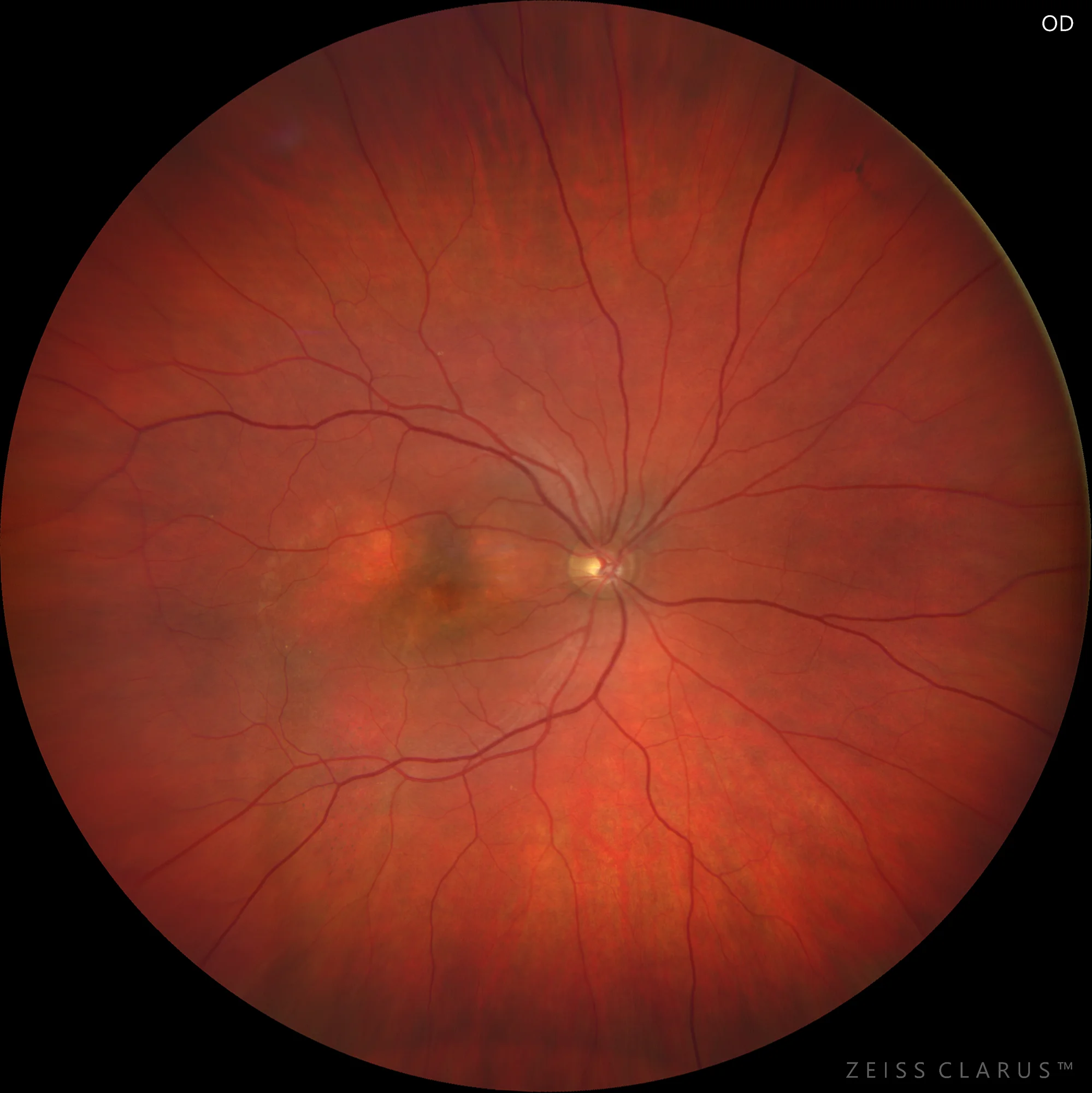
Color retinography showing a reddish fundus with patchy alterations in pigmentation.
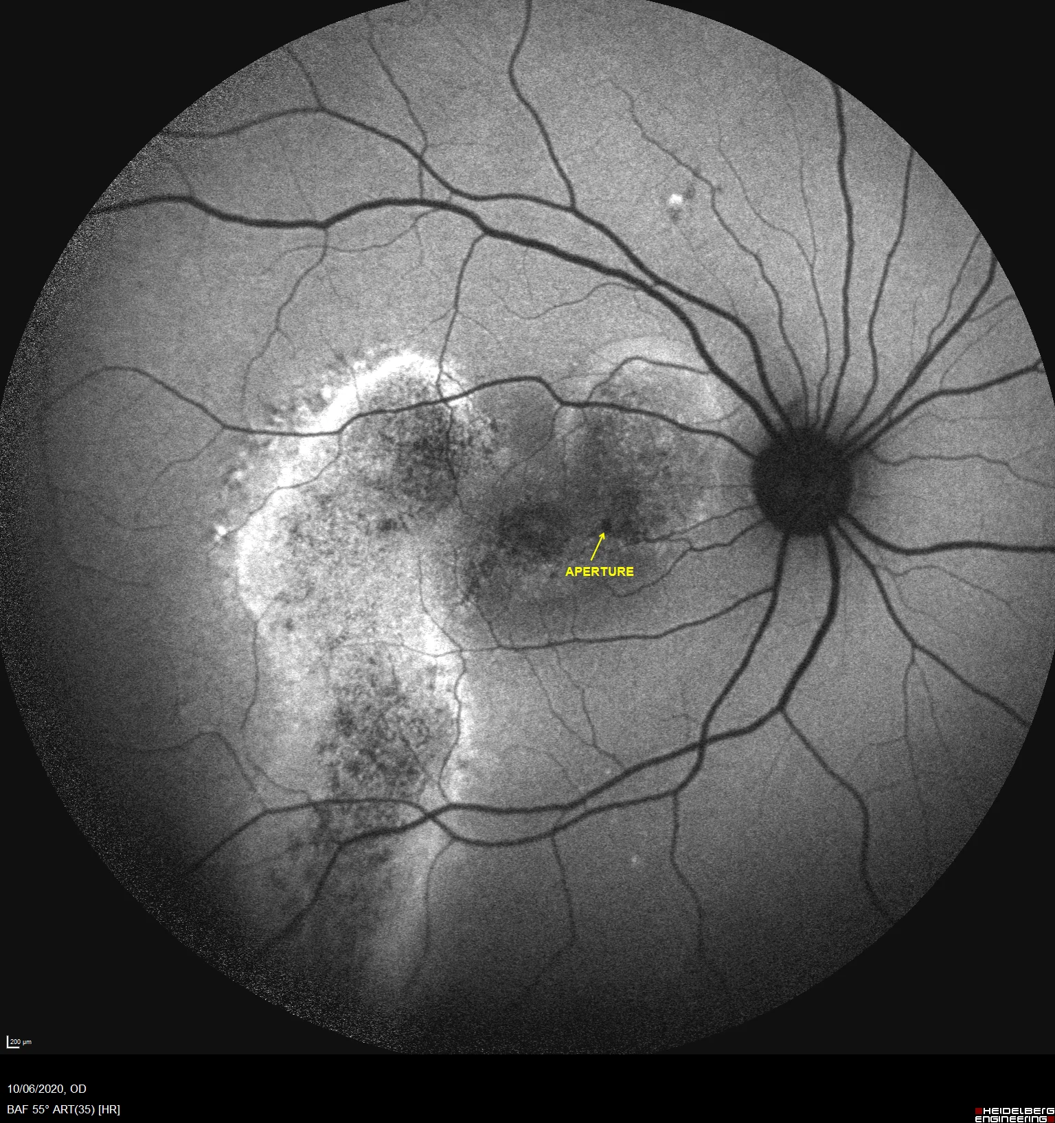
Blue autofluorescence reveals two patches of mottled RPE alteration, with a streaky image from the temporal lesion. In the image, we indicate the presence of a hypoautofluorescent focus that coincides with an image of an RPE opening.
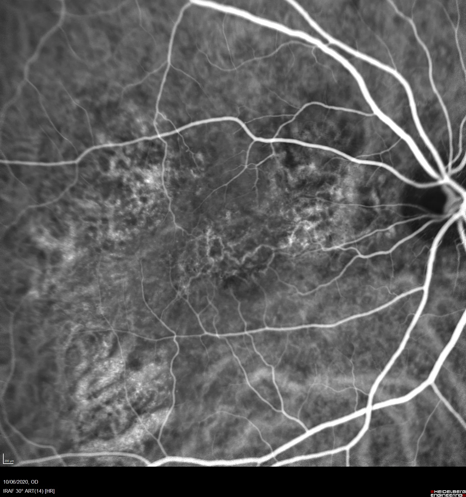
Indocyanine green shows the presence of a reticulated image of fine vessels in the macular area. This vascular network corresponds to the PNV.
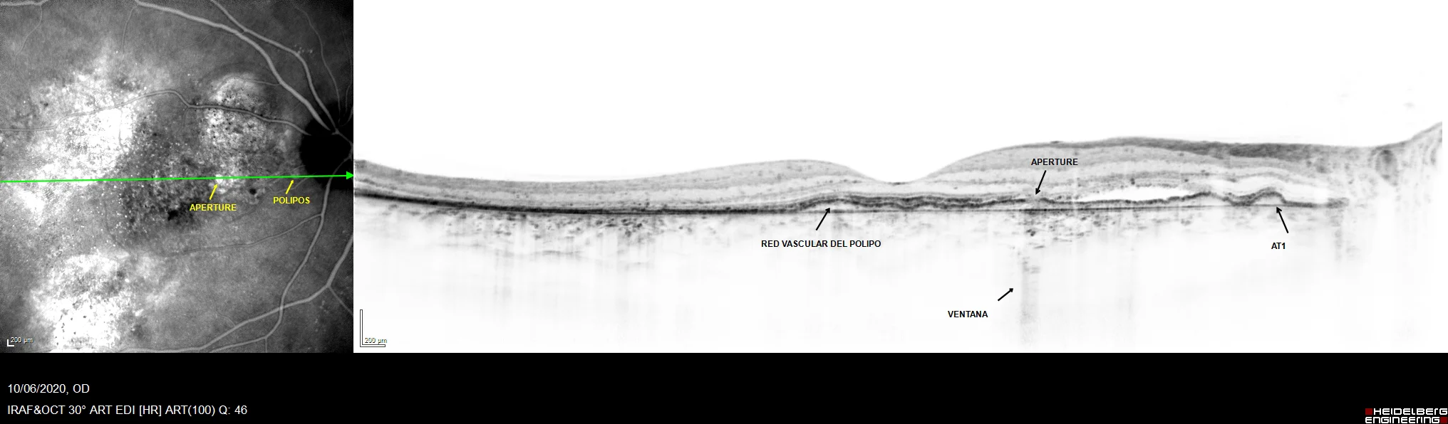
OCT shows an irregular flat elevation of the RPE, with hypo- and hyperreflective spaces within it. This is where the PNV is located. We also highlight the presence of other characteristic findings of pachychoroid disease, such as the window effect of an RPE opening, or the presence of a PED compatible with the presence of type 1 aneurysmal dilations (AT1).
Description
Pachychoroid neovasculopathy (PNV) is a pathological entity characterized by the development of neovessels beneath the retinal pigment epithelium (RPE) in the context of a patient with clinical signs of pachychoroid disease. This condition falls within the spectrum of pachychoroid diseases and is frequently seen associated with central serous chorioretinopathy and polypoidal choroidal vasculopathy.