Pachychoroid peripapillary epitheliopathy
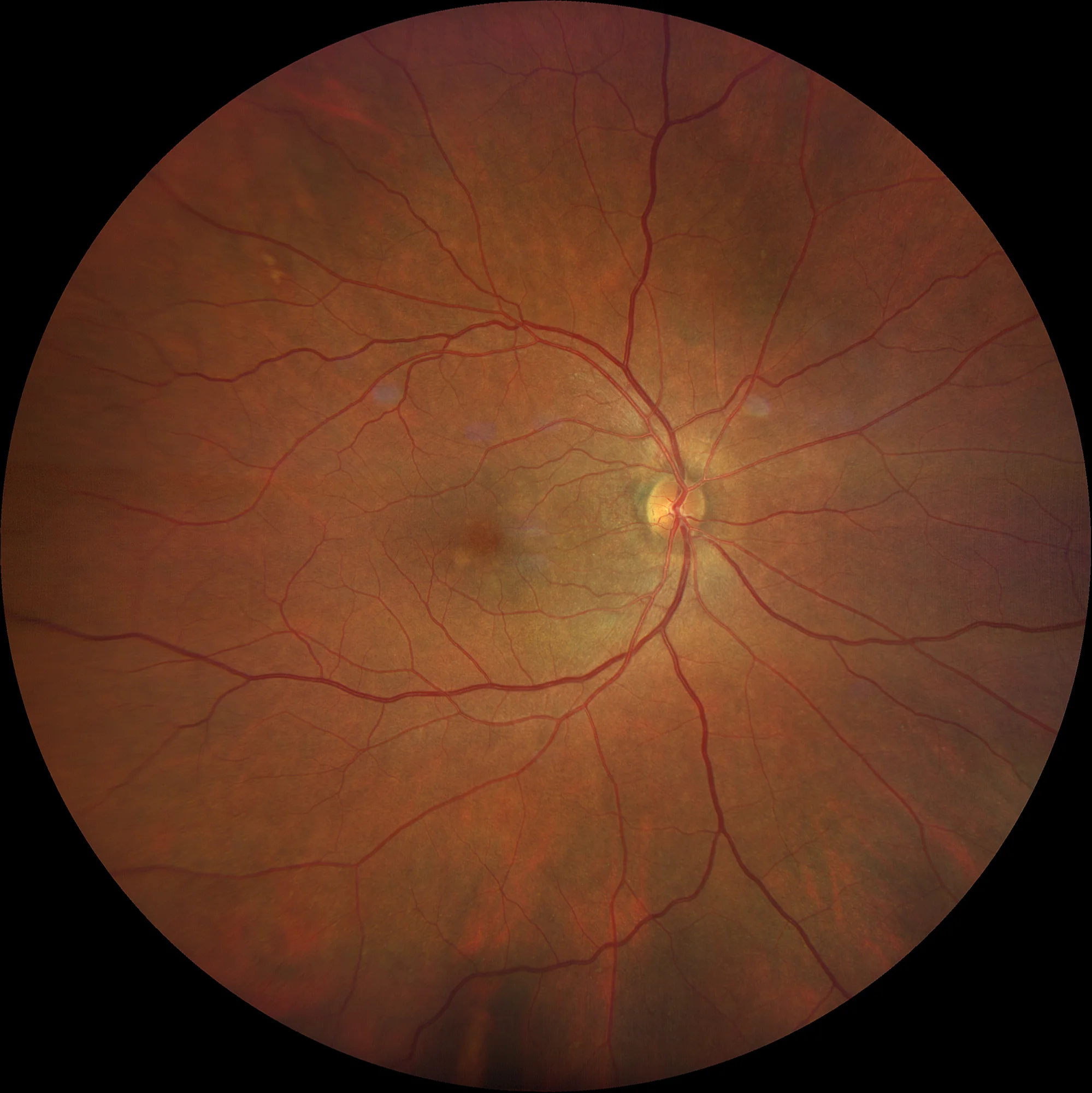
Color retinography showing deterioration and lightening of the peripapillary pigmentation. Some isolated peripheral drusen.
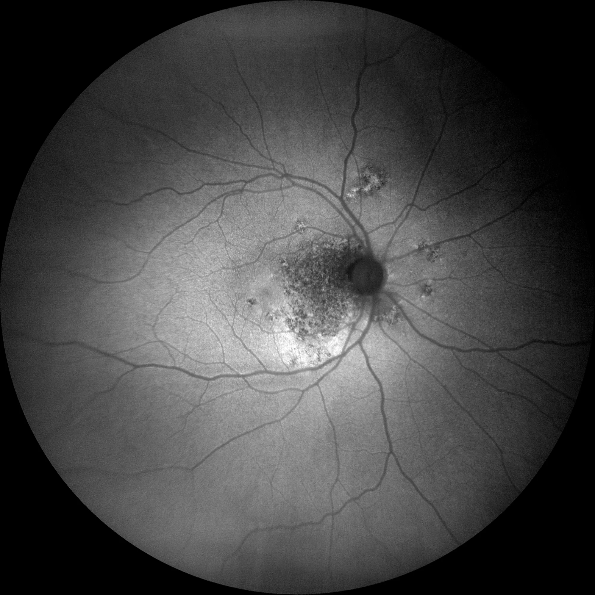
Green autofluorescence: a patchy alteration of the RPE appears, but in a more acute form around the papilla. Hyperautofluorescent border at the lower margin of the lesion.
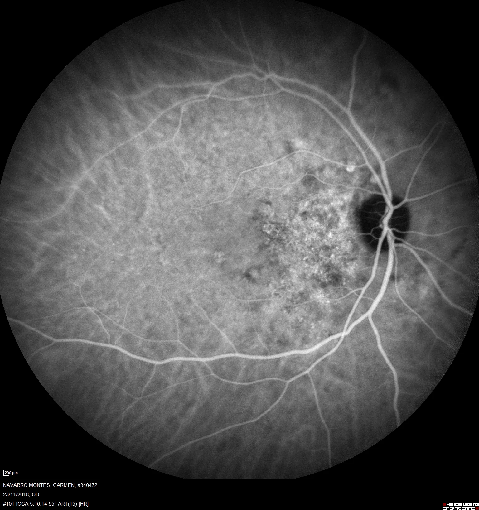
Indocyanine green shows a fine reticular image over the peripapillary patch, suggesting the presence of neovascularization.
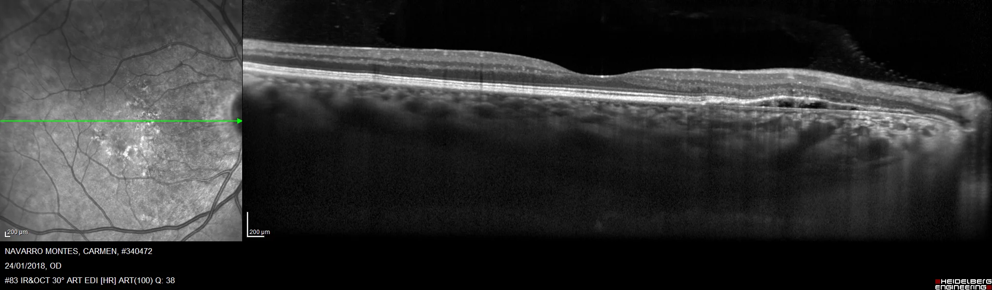
On OCT, an irregular flat elevation of the RPE can be seen. The choroid appears thickened, especially towards the nasal side of the macula.
Description
Pachychoroid peripapillary epitheliopathy (PPE) is a clinical entity characterized by changes in the retinal pigment epithelium (RPE) around the optic nerve, associated with significant choroidal thickening. This condition is seen as part of the spectrum of pachychoroid diseases, which includes central serous chorioretinopathy and polypoidal chorioretinopathy. Patients with PPE may present with subretinal fluid, pigmented epithelial atrophies, and a congested optic disc.