< back
Pachychoroid pigment epitheliopathy
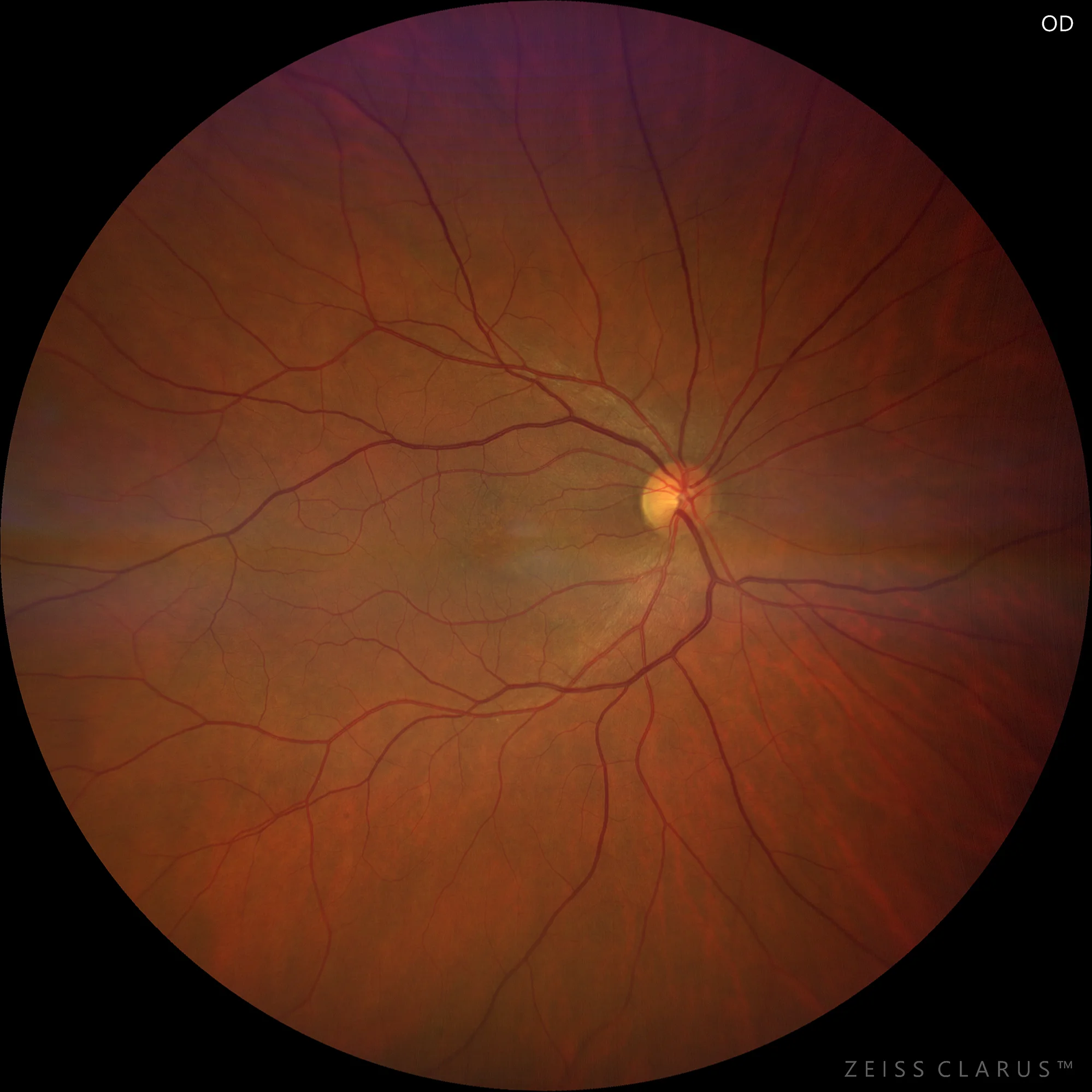
Color retinography showing a mottling of pigmentary alterations in the macula and in the region inferonasal to the macula.
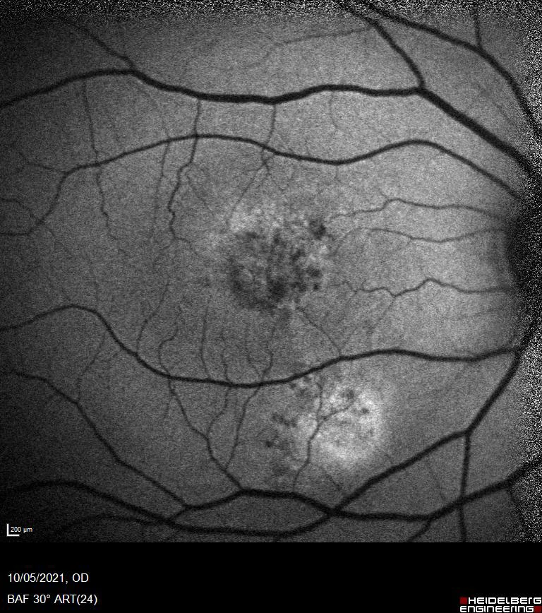
Green autofluorescence: Hypoautofluorescent mottling is observed secondary to epitheliopathy.
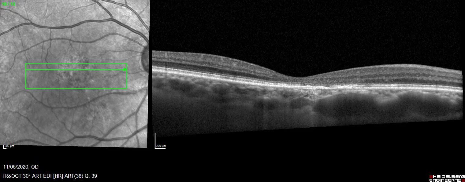
OCT shows retinal thinning due to atrophy of the outer retina, ellipsoids, and outer nuclear areas. The presence of prominent choroidal vessels close to the inner choroid is notable.
Description
Pachychoroid pigment epitheliopathy (PPE) is a clinical entity characterized by the presence of changes in the retinal pigment epithelium (RPE) associated with an abnormally thickened choroid. These patients do not present subretinal fluid, but share all their pathophysiology with central serous chorioretinopathy.