Casos
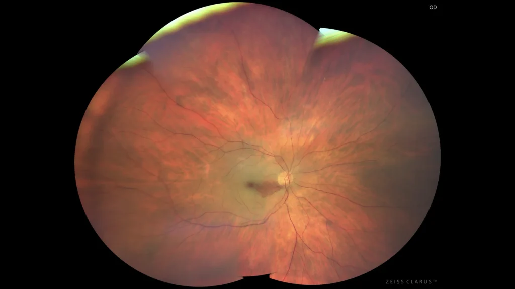
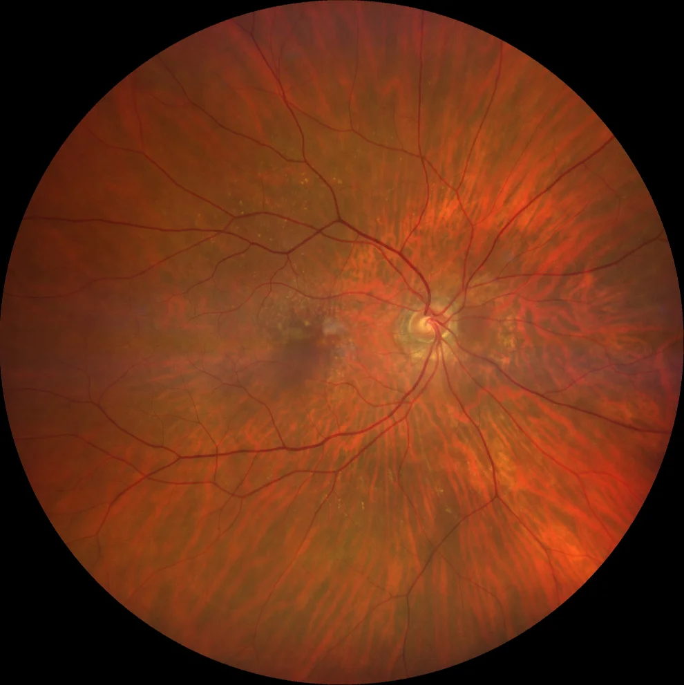
Central retinal artery occlusion
Central retinal artery occlusion (CRAO) is an ophthalmic emergency usually caused by an embolus blocking the central retinal artery, leading to sudden
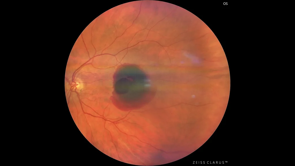
Retinal arterial macroaneurysm
Retinal arterial macroaneurysm (MAR) is an abnormal dilation of the retinal arteries, most common in older women and often associated with hypertensio
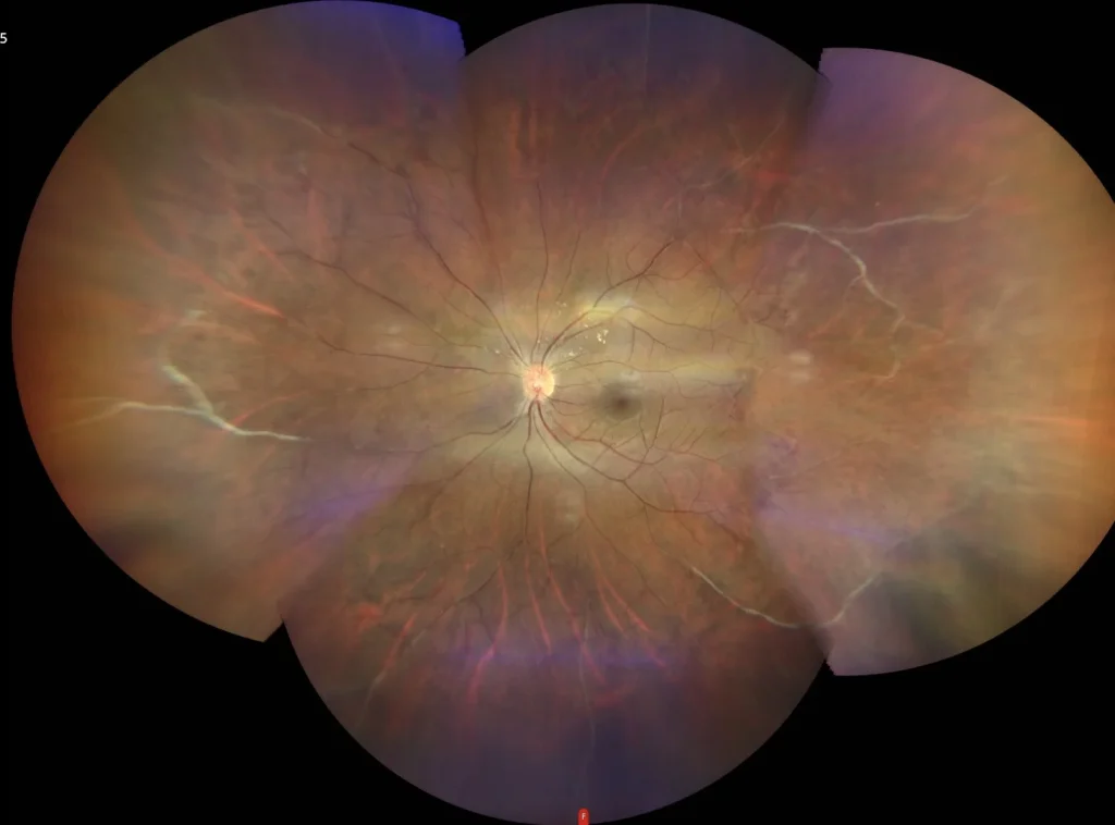
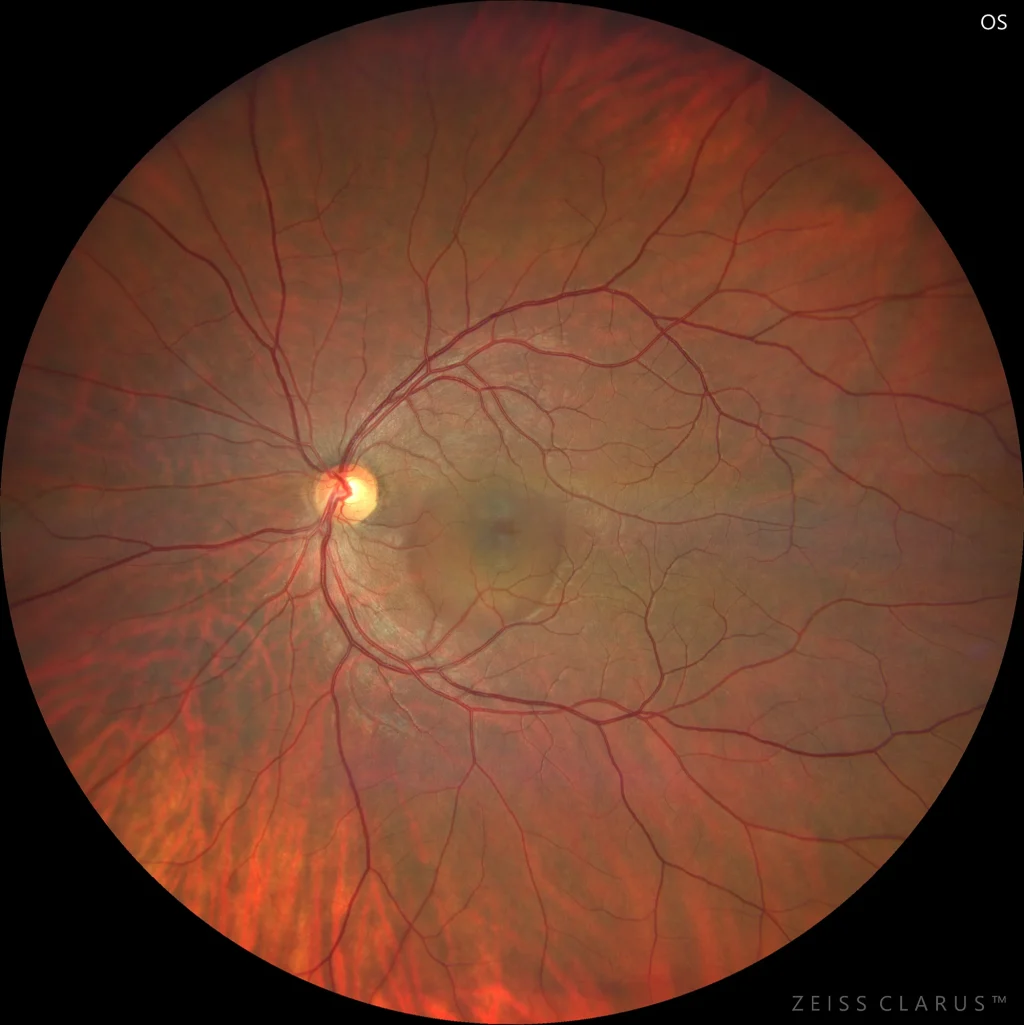
Eales disease
Eales disease is an idiopathic occlusive peripheral periphlebitis that primarily affects young men, particularly in the Indian subcontinent. It is cha
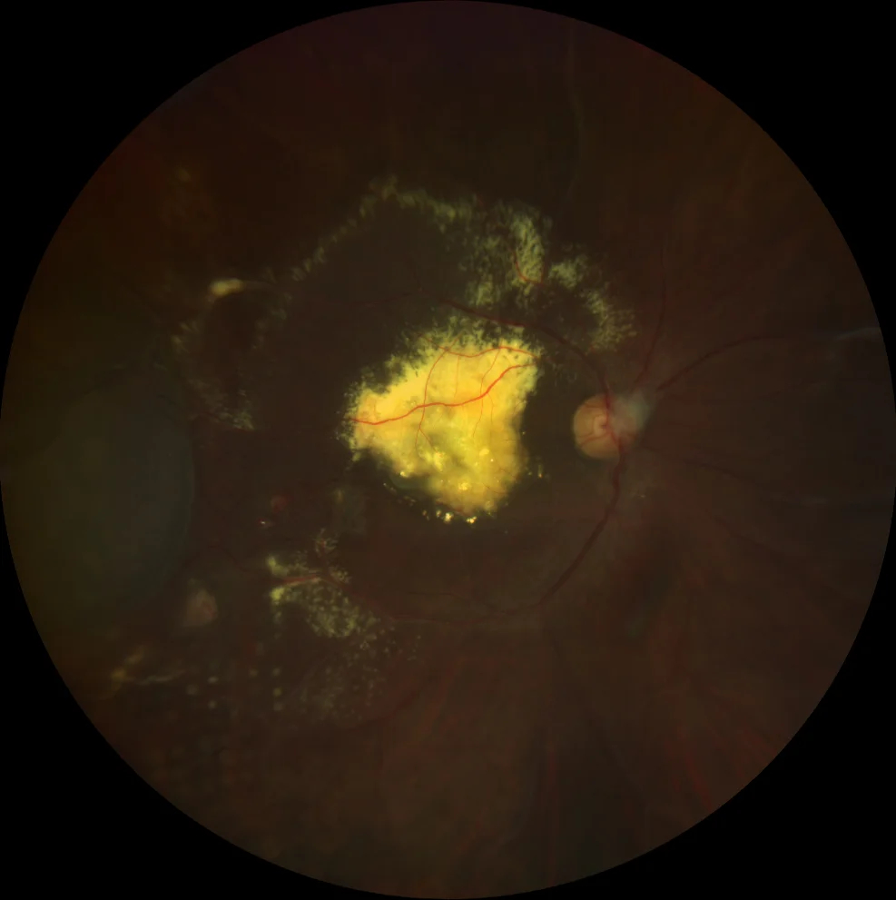
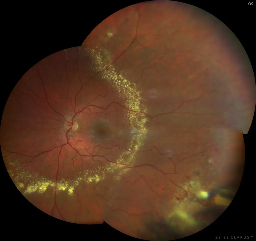
Coats disease
Coats disease is an idiopathic retinal vasculopathy characterized by telangiectasias and retinal aneurysms and intra- and subretinal exudation. It typ
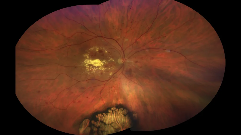
Coats disease
Coats disease is a non-inherited condition characterized by the presence of unilateral retinal telangiectasias, exudation, and exudative retinal detac
Diabetic macular edema
Diabetic macular edema (DME) is evident on optical coherence tomography as macular thickening with hyporeflectivity due to intraretinal fluid accumula
Acute central serous chorioretinopathy
Acute central serous chorioretinopathy. Acute central serous chorioretinopathy (CSCR) is an ocular pathology characterized by the accumulation of ser
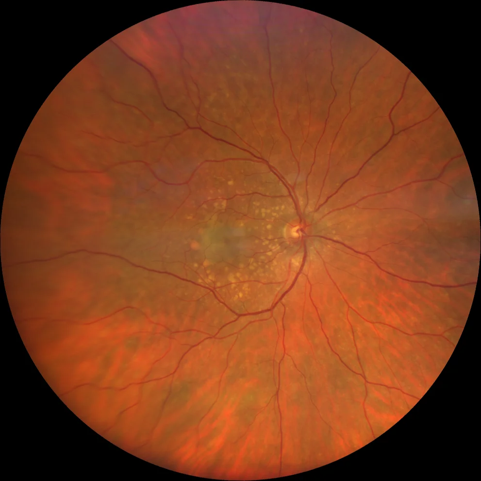
Subretinal drusenoid deposits
76-year-old woman with minimal vision loss in her right eye who comes for a check-up.
AVMC OD 20/30 OI 20/20
Reticular pseudodrusen and soft druse
Soft drusen and serous PED
73-year-old woman with vision loss and metamorphopsia in the right eye of 1 month\'s duration
AVMC OD 20/50 OI 20/20
Soft drusen in both eyes. Serou