Casos
Drusenoid detachment of pigmented epithelium
Drusenoid detachment of the pigmented epithelium. The presence of large soft duras causes them to merge, giving rise to an image of detachment of the
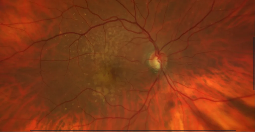
geographic atrophy
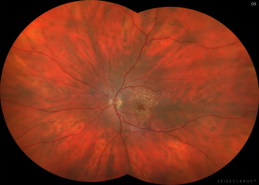
78-year-old woman who comes to the consultation for a check-up.
VA in LE is 20/20. An area of extrafoveal geographic atrophy is observed in the fundu
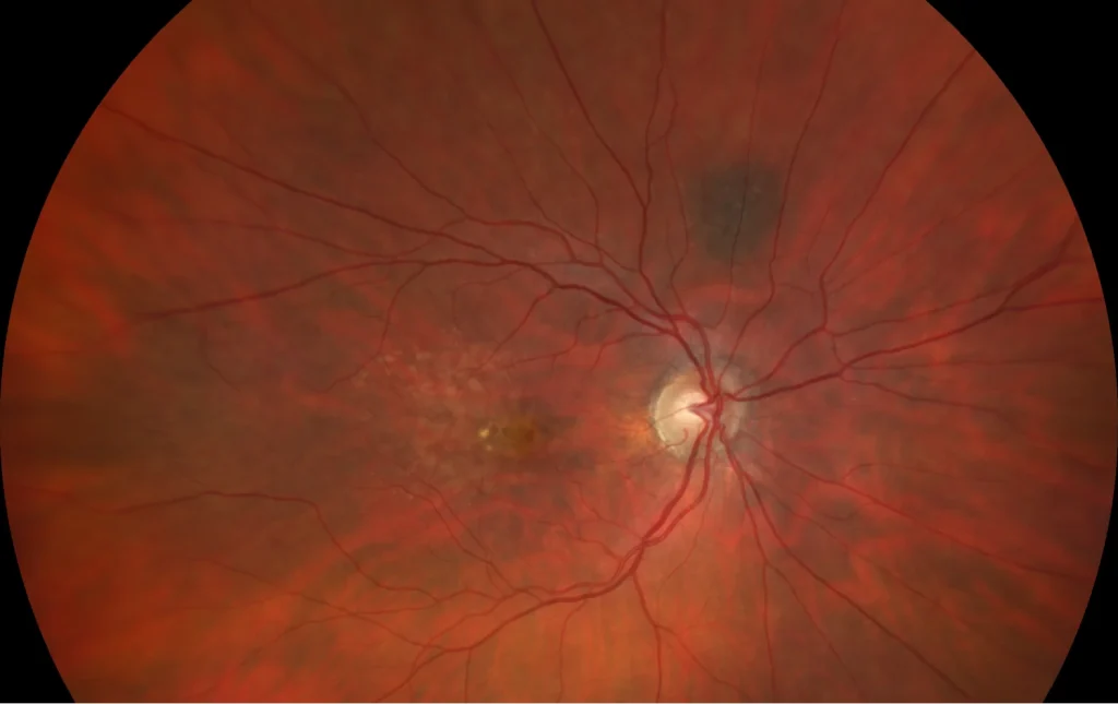
Neovascular membrane type 3
Neovascular age-related macular degeneration (AMD) accounts for approximately 1 in 10 cases of AMD. In particular, in type 3 neovascularization, neove
Macular neovascularization type 3
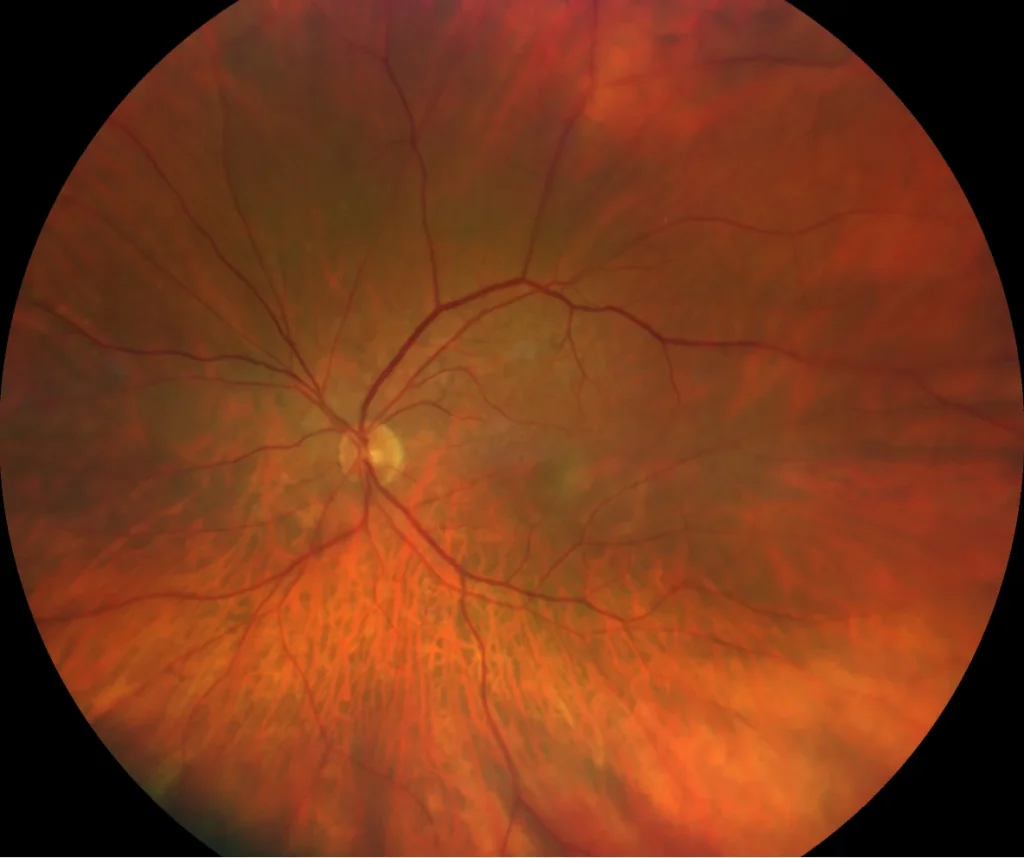
76-year-old woman who comes due to loss of vision in her left eye.
VA in the RE is 20/20 and in the LE 20/50. She is pseudophakic in both eyes.
The
Retinal Angiomatous Proliferation Stage 1
Characteristics:
The current classification of neovascular Age-Related Macular Degeneration (AMD) is based on the findings of Optical Coherence Tomog
Macular neovascularization type 2
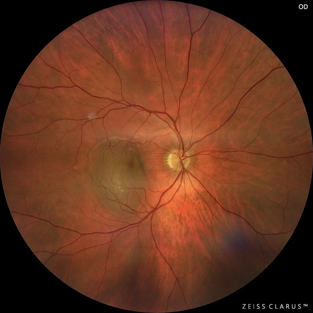
85-year-old woman who comes due to loss of vision in her left eye.
VA in RE is 20/25 and in LE 20/50. He has 2+ nuclear cataracts in both eyes.
The
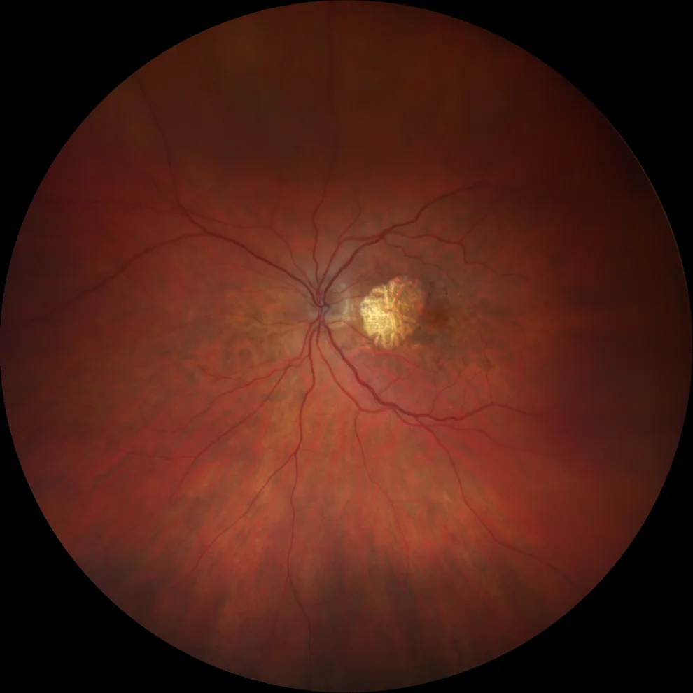
Aneurysmal type 1 neovascular membrane
This is a subtype of exudative age-related macular degeneration characterized by a growth of the neovascular complex beneath the pigmented epithelium.

Type 1 macular neovascularization with aneurysmal dilatations
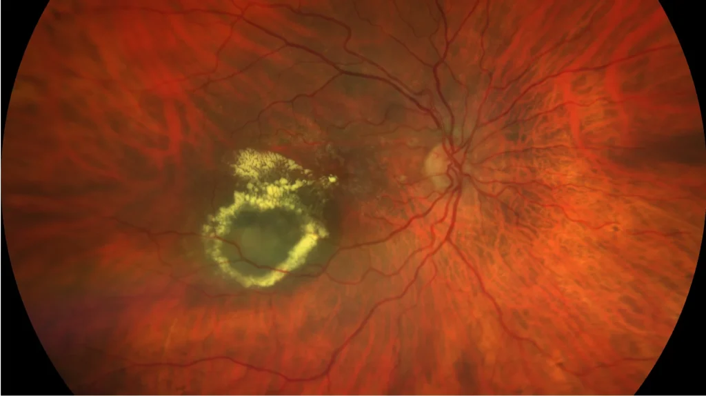
75-year-old woman who comes due to loss of vision in her right eye.
VA in OD is 20/80. It is phakic.
The fundus reveals multiple hard exudates in t
Aneurysmal neovascularization type 1
Aneurysmal neovascularization type 1, also known as polypoidal choroidal vasculopathy (PCV), is a condition characterized by the presence of sub-pigme