Casos
Myopic macular hemorrhage
A 47-year-old male comes to the doctor due to sudden loss of vision in his right eye.
AV OD 20/40 with -9.00 -4.25 x 160º.
Fundus examination revea
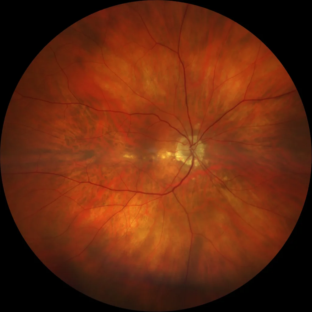
Vanishing hemorrhage
Evanescent hemorrhages in myopes are usually subretinal hemorrhages (sometimes intraretinal) that occur during the development or expansion of a lacqu
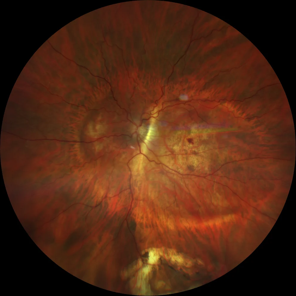
Lacquer Streak
Lacquer striae are considered to be healed mechanical tears of the choriocapillaris-Bruch\'s membrane complex. They appear as single or multiple, irre
Lacquer Streak
Lacquer striae are considered to be healed mechanical tears of the choriocapillaris-Bruch\'s membrane complex. They appear as single or multiple, irre
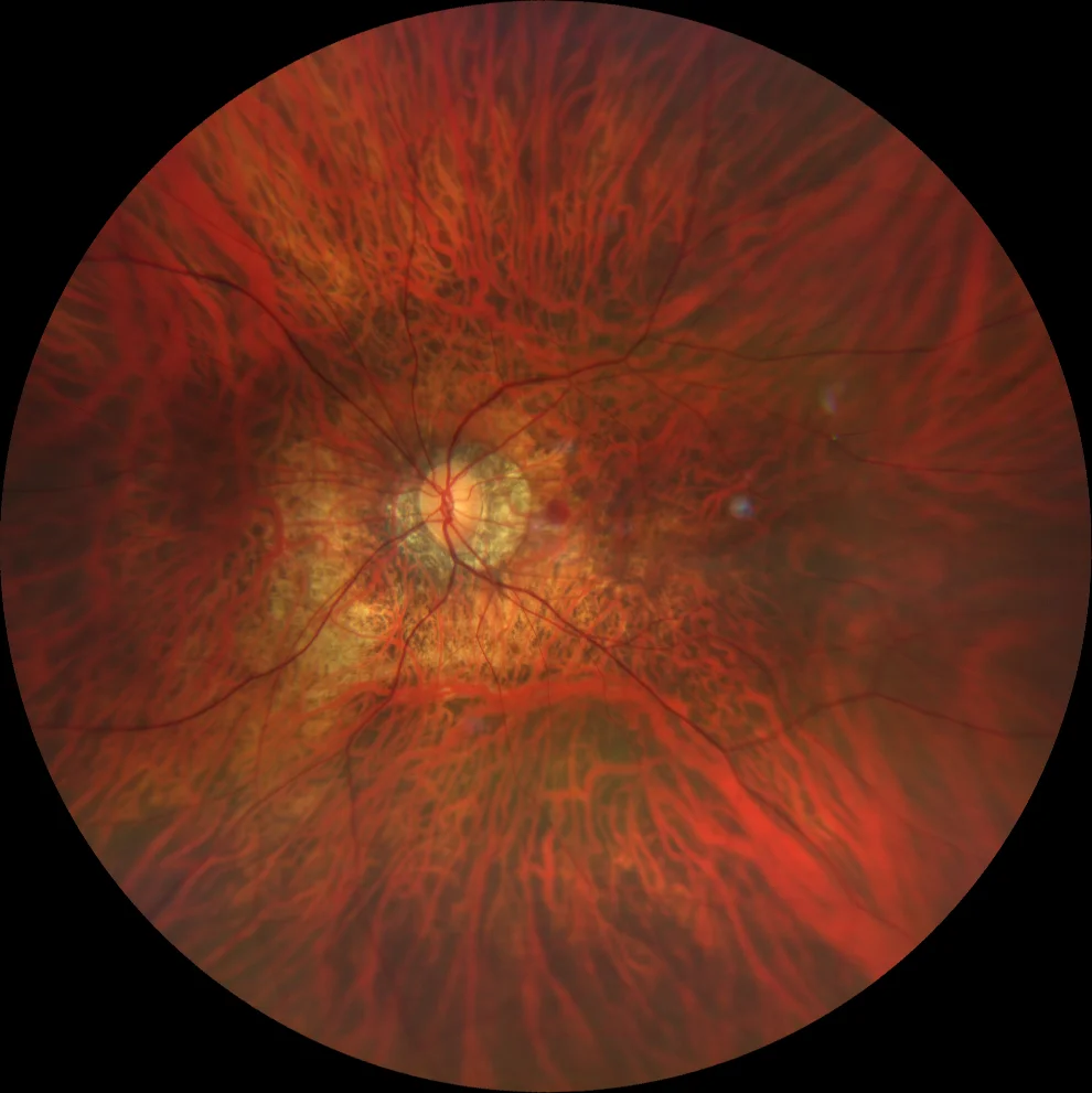
Posterior Staphyloma
Posterior staphyloma is the most characteristic sign and the main marker of pathological myopia. Spaide defined it as an evagination or protrusion of
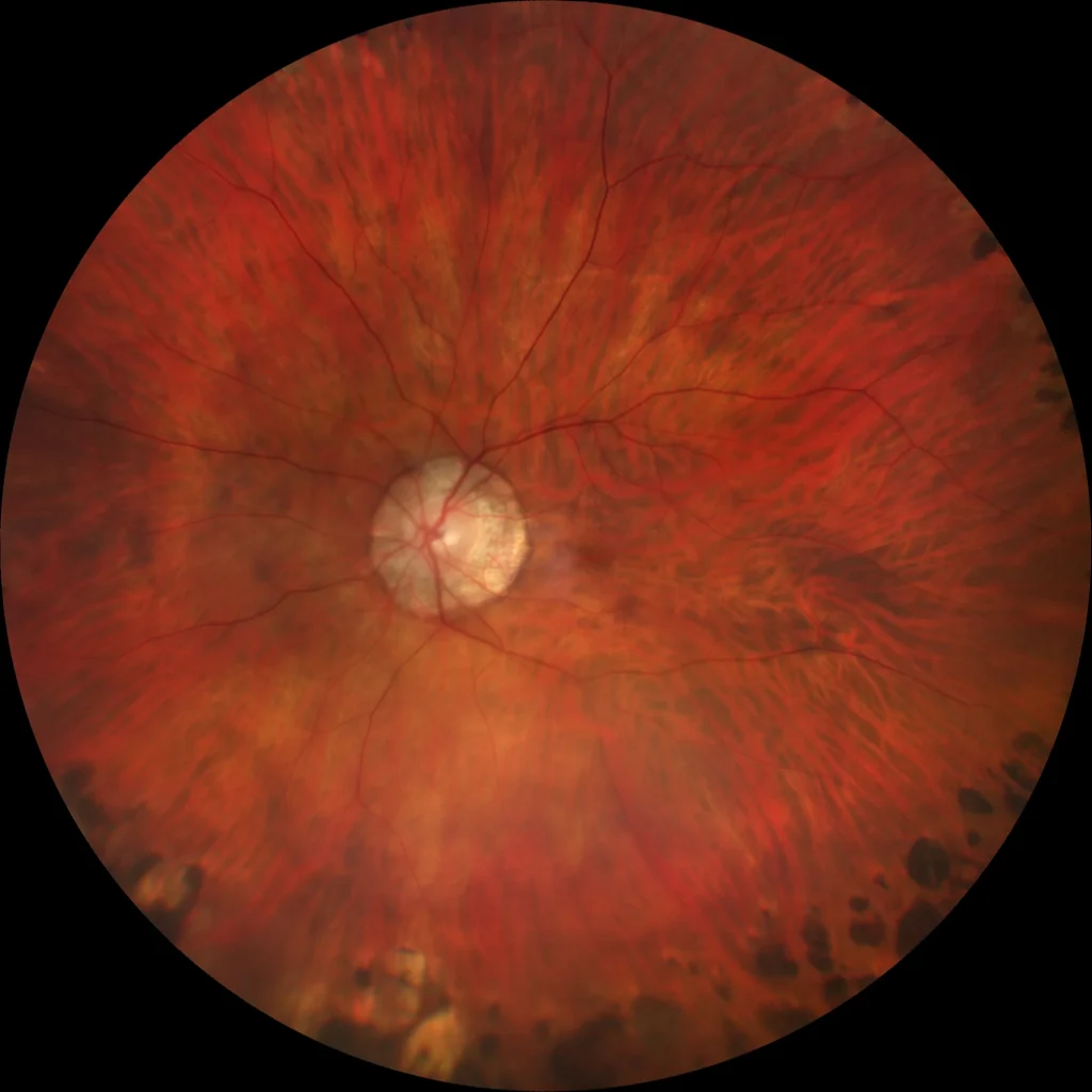
Septated staphyloma with myopic macular neovascularization
A 66-year-old male presents with a 2-week history of central scotoma in the left eye. His ophthalmic history includes pathological myopia in both eyes
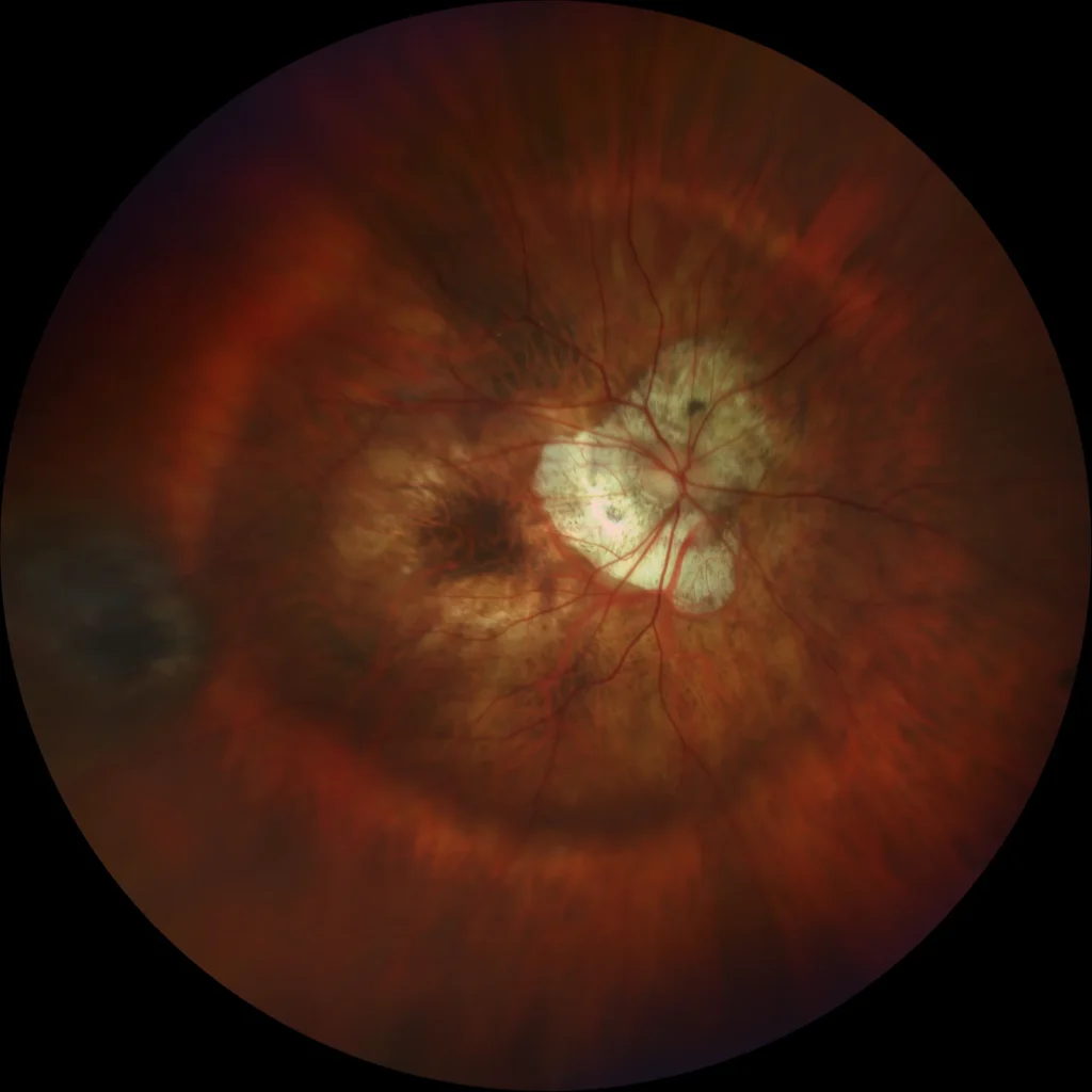
Posterior Staphyloma
Posterior staphyloma is the most characteristic sign and the main marker of pathological myopia. Spaide defined it as an evagination or protrusion of
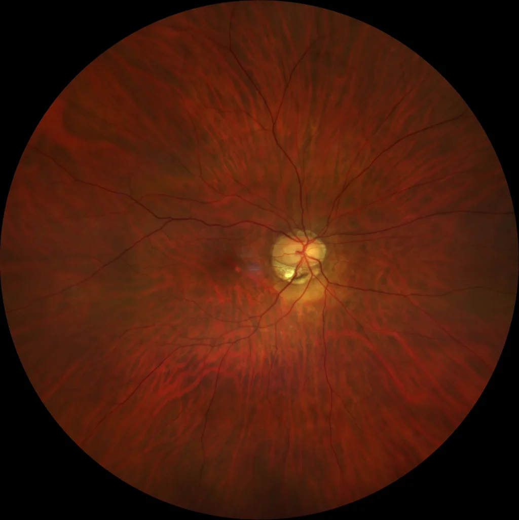
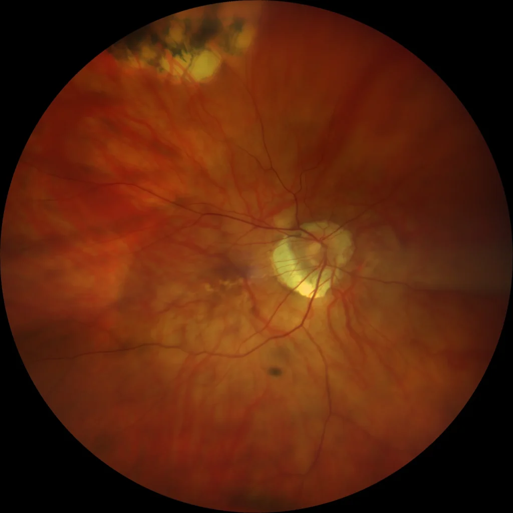
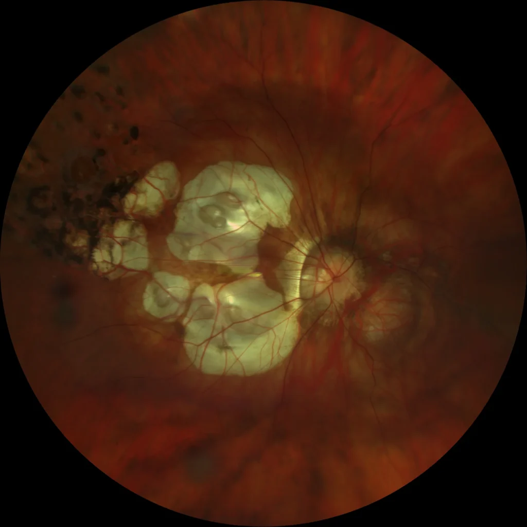
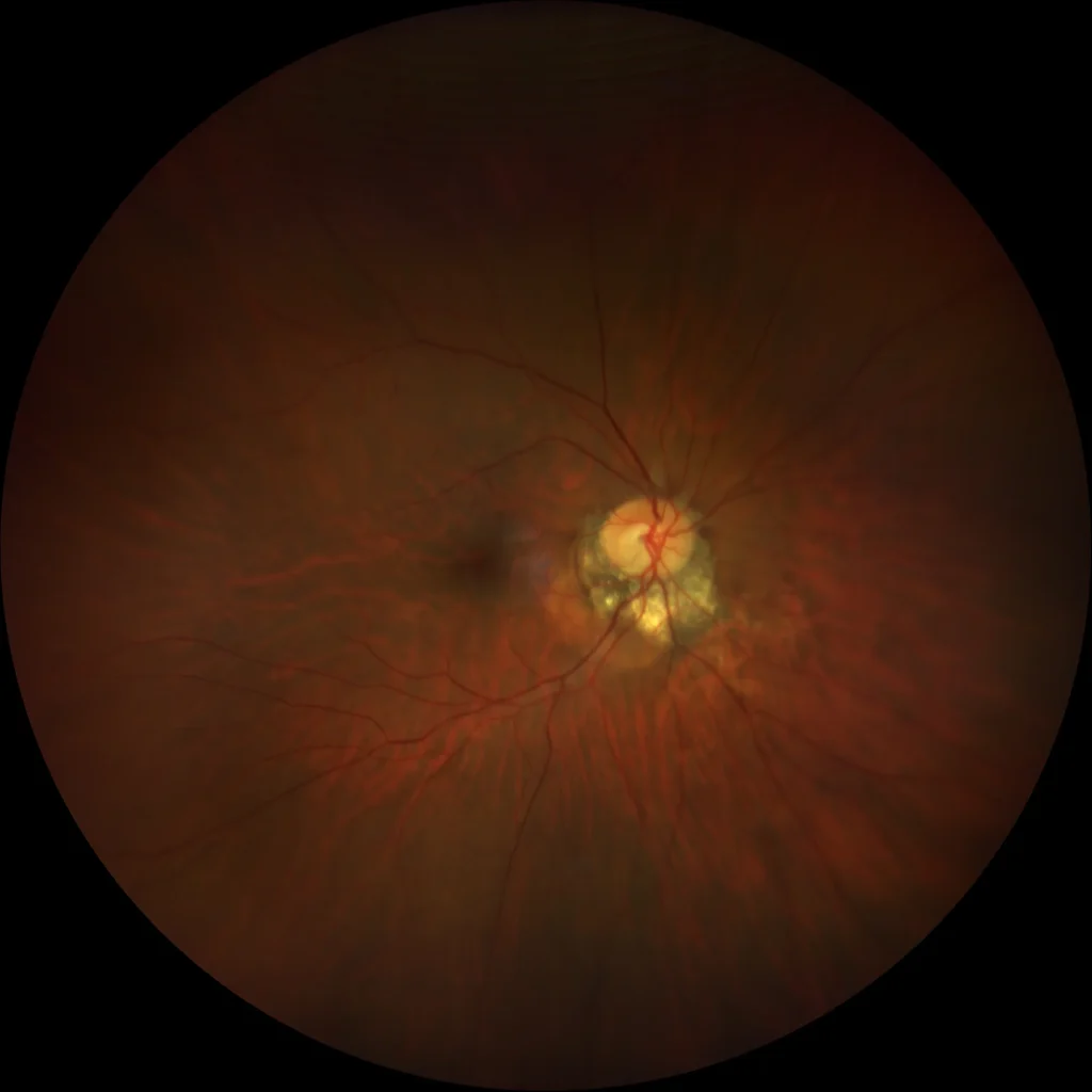
Peripapillary Intrachoroidal Cavitation
Well-defined yellow-orange lesion, located at the outer edge of the myopic cone (generally at the lower outer edge of the peripapillary gamma atrophy)
Peripapillary Intrachoroidal Cavitation
Well-defined yellow-orange lesion, located at the outer edge of the myopic cone (generally at the lower outer edge of the peripapillary gamma atrophy)