Papillary drusen and angioid streaks

Color retinography (CLARUS 500, Zeiss): drusen visible at the papillary level. Angioid streaks arranged around the papilla and in a radial manner. “Orange peel” image temporal to the macular area.
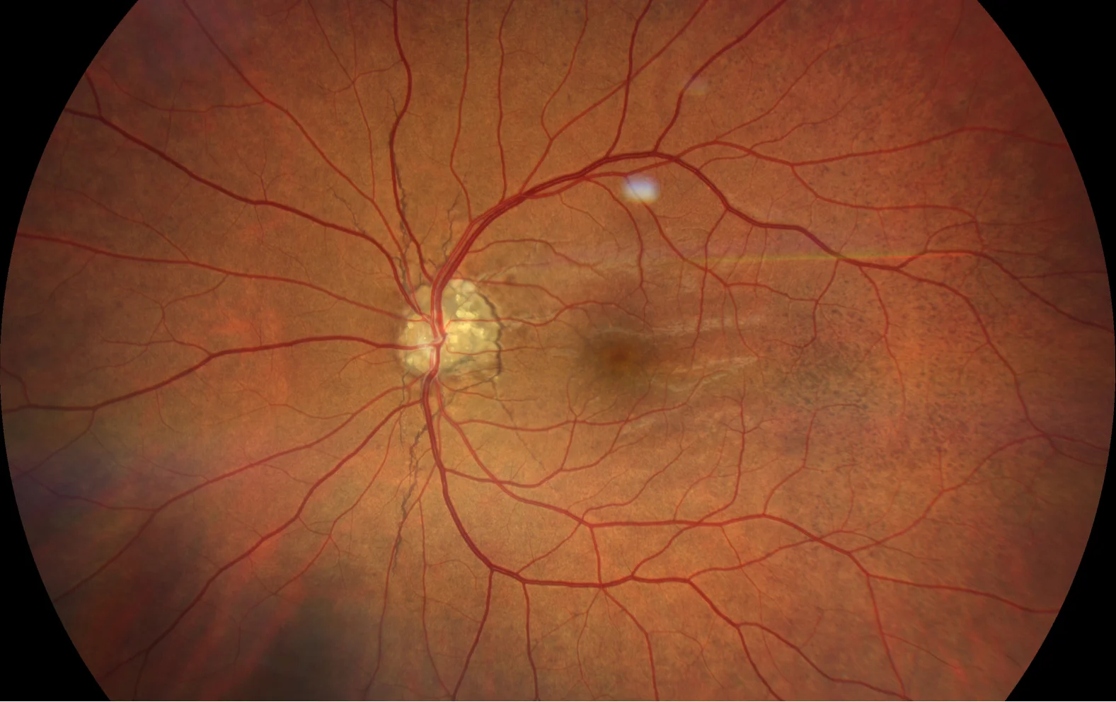
Color retinography (CLARUS 500, Zeiss): drusen visible at the papillary level. Angioid streaks arranged around the papilla and in a radial manner. “Orange peel” image temporal to the macular area.
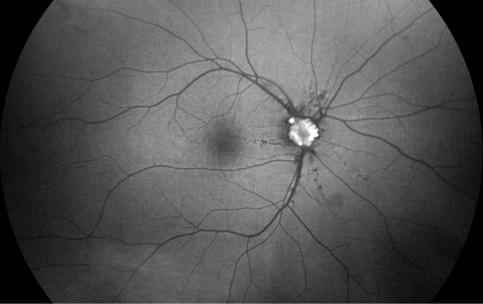
Autofluorescence (CLARUS 500, Zeiss): marked prepapillary hyperautofluorescence with linear hypoautofluorescence of the angioid streaks
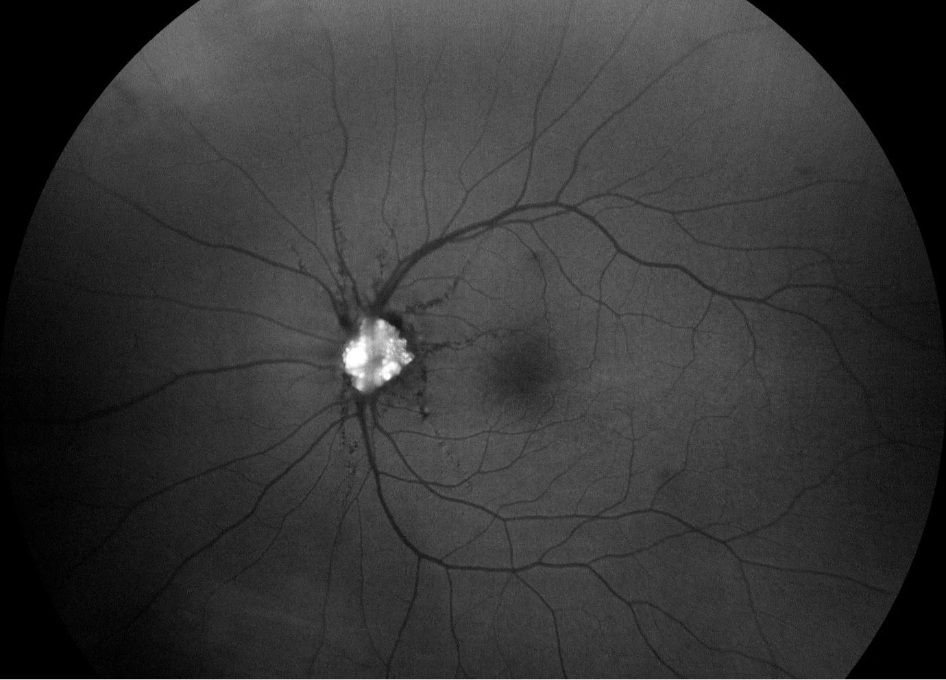
Autofluorescence (CLARUS 500, Zeiss): marked prepapillary hyperautofluorescence with linear hypoautofluorescence of the angioid streaks
Description
Papillary drusen and angioid streaks . Papillary drusen are hyaline formations measuring 5 to 1,000 microns in diameter located in front of the cribriform plate. They may be visible or hidden. They usually cause campimetric alterations secondary to axonal compression, but maintain normal visual acuity. The differential diagnosis should be made with papilledema. Angioid streaks may coexist in pathologies such as pseudoxanthoma elasticum.