Pattern Dystrophy – Butterfly Wing
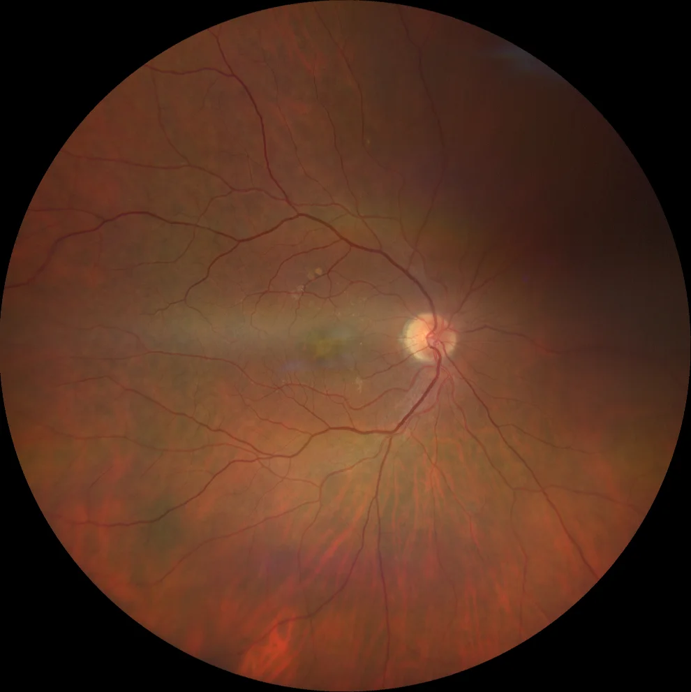
Retinography (Clarus 700, Zeiss): yellowish and pigmented lesions in a radial arrangement in the left eye (A2) and in a more central location in the right eye (A1).
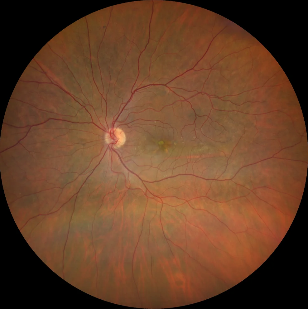
Retinography (Clarus 700, Zeiss): yellowish and pigmented lesions in a radial arrangement in the left eye (A2) and in a more central location in the right eye (A1).
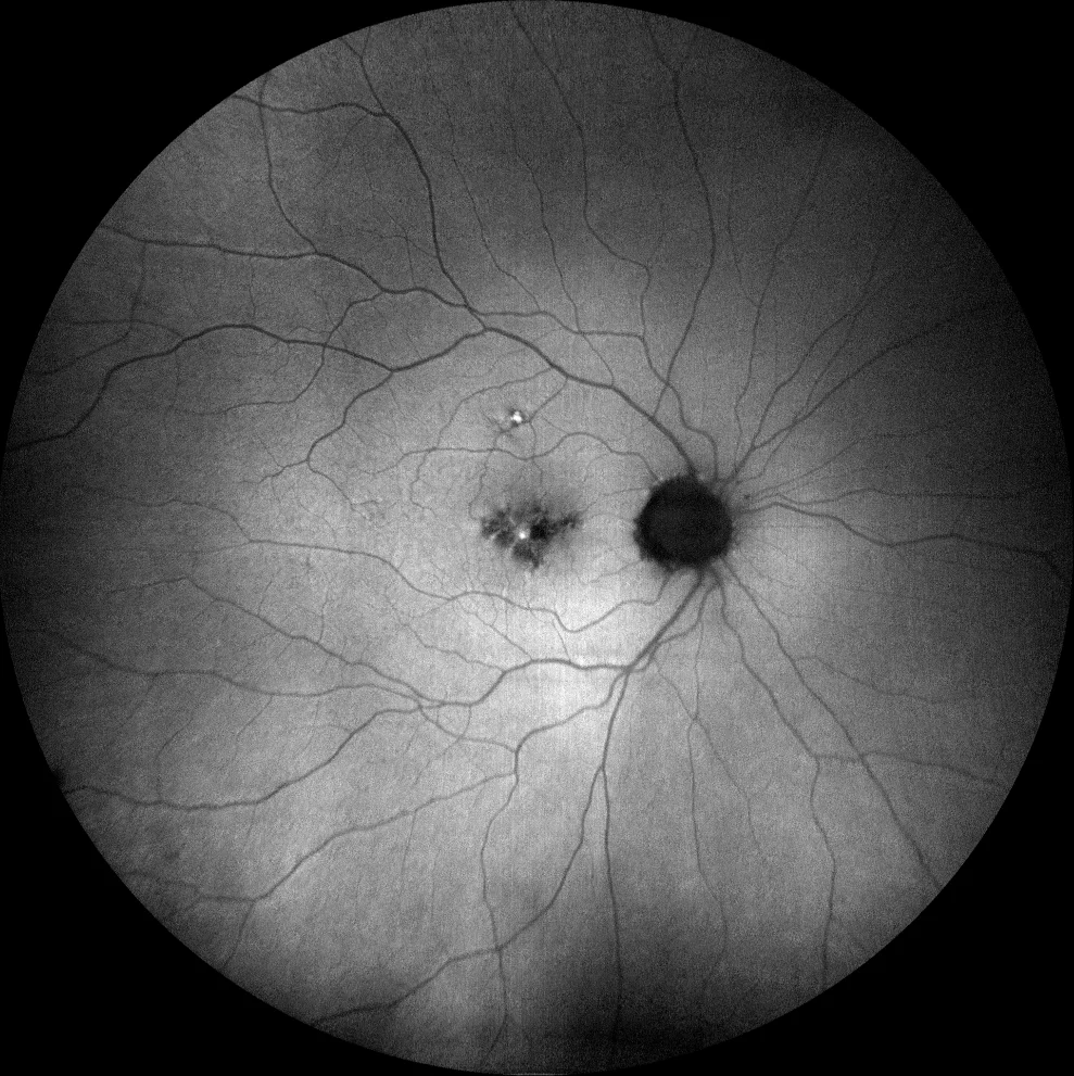
Green autofluorescence (Clarus 700, Zeiss): The lesions have a hyperautofluorescent component corresponding to the vitelliform material and a hypofluorescent component corresponding to the pigment deposit (B1, B2).
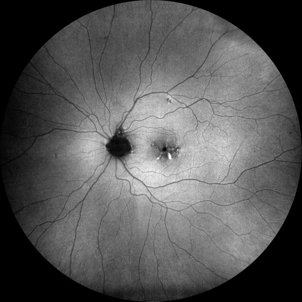
Green autofluorescence (Clarus 700, Zeiss): The lesions have a hyperautofluorescent component corresponding to the vitelliform material and a hypofluorescent component corresponding to the pigment deposit (B1, B2).
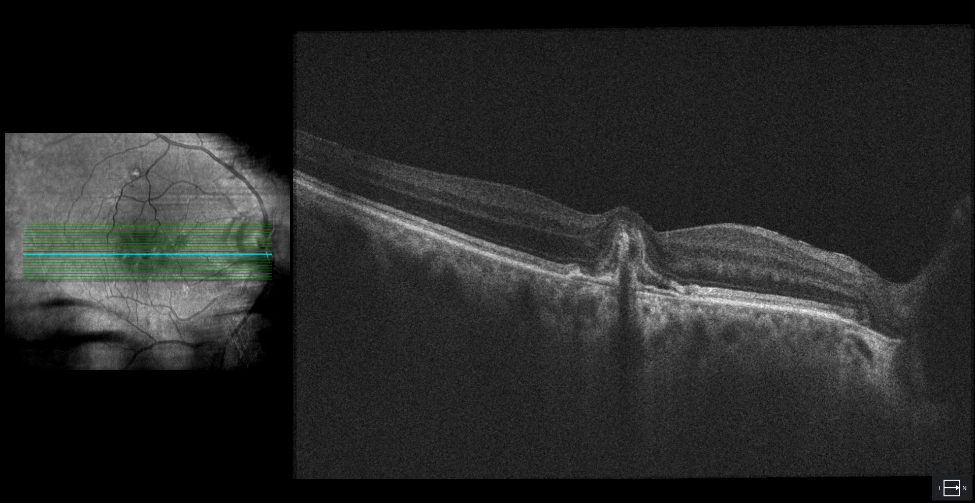
OCT (Cirrus 5000-HD, Zeiss) and OCT-angiography (Cirrus 5000-HD, Angioplex, Zeiss): the right eye shows a subretinal hyperreflective material at foveal level and an underlying double layer sign (C1). This strongly suggests the presence of NVM, which is confirmed by OCT-angiography (C2). The left eye shows outer retinal atrophy and RPE at foveal level (C3).
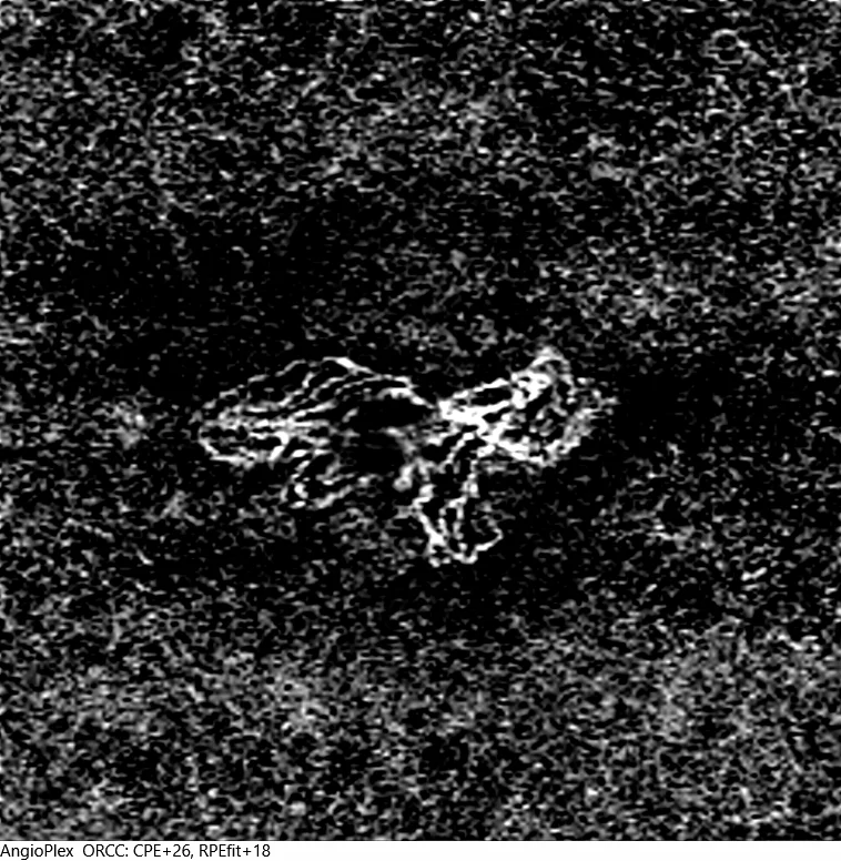
OCT (Cirrus 5000-HD, Zeiss) and OCT-angiography (Cirrus 5000-HD, Angioplex, Zeiss): the right eye shows a subretinal hyperreflective material at foveal level and an underlying double layer sign (C1). This strongly suggests the presence of NVM, which is confirmed by OCT-angiography (C2). The left eye shows outer retinal atrophy and RPE at foveal level (C3).
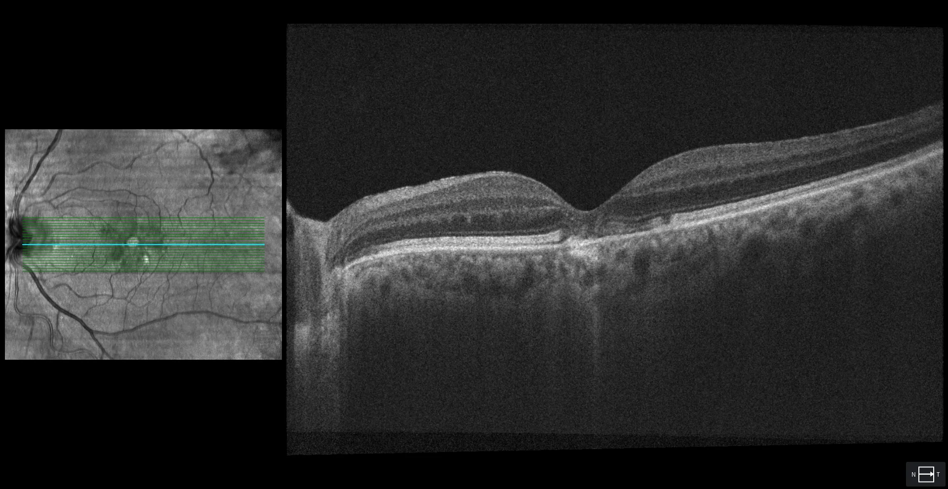
OCT (Cirrus 5000-HD, Zeiss) and OCT-angiography (Cirrus 5000-HD, Angioplex, Zeiss): the right eye shows a subretinal hyperreflective material at foveal level and an underlying double layer sign (C1). This strongly suggests the presence of NVM, which is confirmed by OCT-angiography (C2). The left eye shows outer retinal atrophy and RPE at foveal level (C3).
Description
A 77-year-old male diagnosed with pattern dystrophy – butterfly wing presents with vision loss in the right eye.
VA OD 20/50 OS 20/30.
Fundus examination shows yellowish lesions and pigment deposits in the macula of both eyes. Autofluorescence of the left eye reveals a hyper-hypoautofluorescent pattern in butterfly wing or cartwheel shape typical of this condition; in the right eye, hyper- and hypoautofluorescent lesions are also observed but without the typical pattern. OCT of the left eye shows atrophy of the outer retina and RPE at the foveal level. OCT of the right eye shows hyperreflective subretinal material and the double layer sign (RPE and Bruch’s membrane) suggesting the presence of choroidal neovascularization (CNV), which is confirmed by angio-OCT.