Pattern Dystrophy – Multifocal Pattern
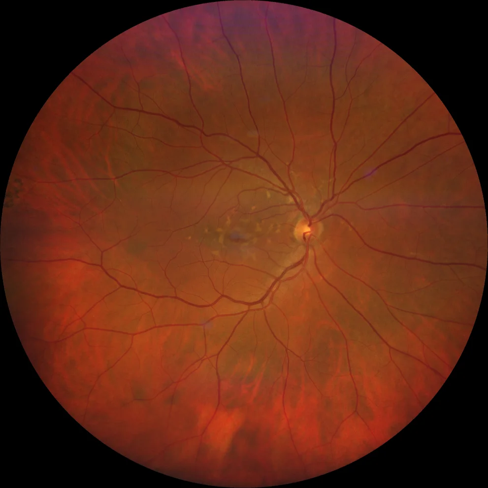
Retinography (Clarus 700, Zeiss): multiple yellowish deposits in the macula and peripapillary area (A1, A2).
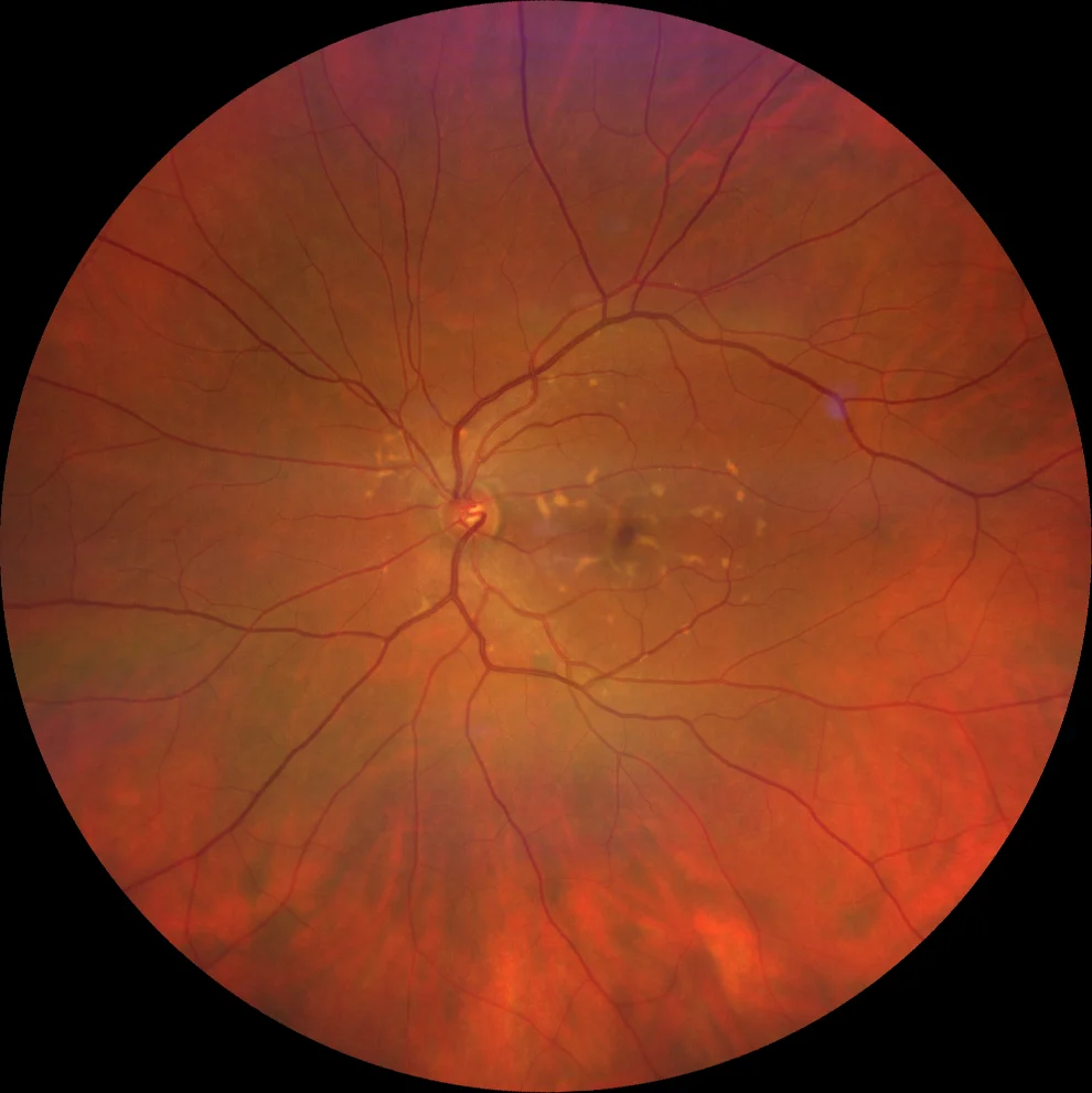
Retinography (Clarus 700, Zeiss): multiple yellowish deposits in the macula and peripapillary area (A1, A2).
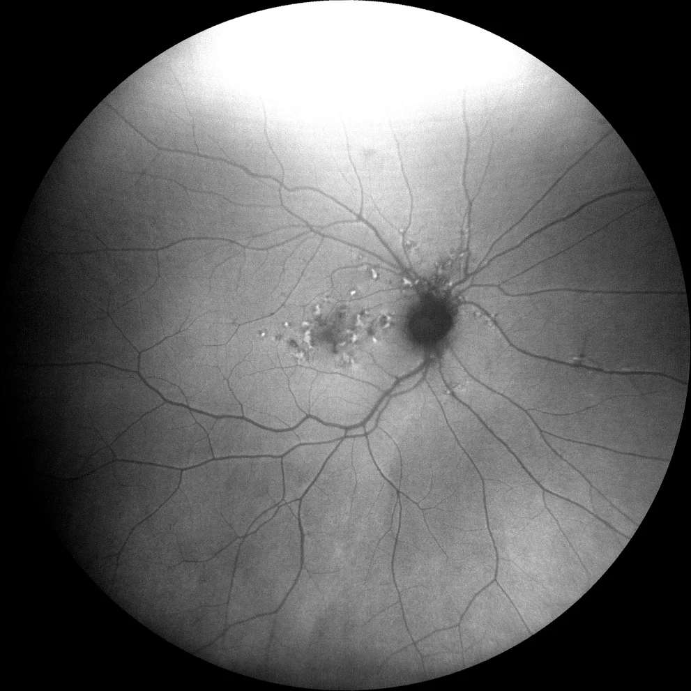
Green autofluorescence (Clarus 700, Zeiss): the deposits are hyperautofluorescent, indicating that they are vitelliform material (lipofuscin). In addition, areas of hypoautofluorescence due to RPE involvement are observed (B1, B2). In the right eye, it can be seen how these areas, unlike what occurs in Stargardt's disease, affect the peripapillary region (B1).
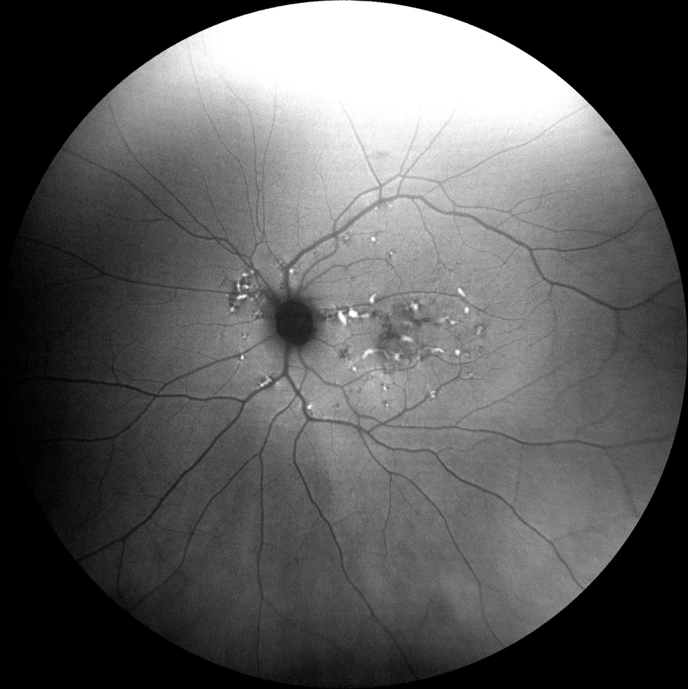
Green autofluorescence (Clarus 700, Zeiss): the deposits are hyperautofluorescent, indicating that they are vitelliform material (lipofuscin). In addition, areas of hypoautofluorescence due to RPE involvement are observed (B1, B2). In the right eye, it can be seen how these areas, unlike what occurs in Stargardt's disease, affect the peripapillary region (B1).
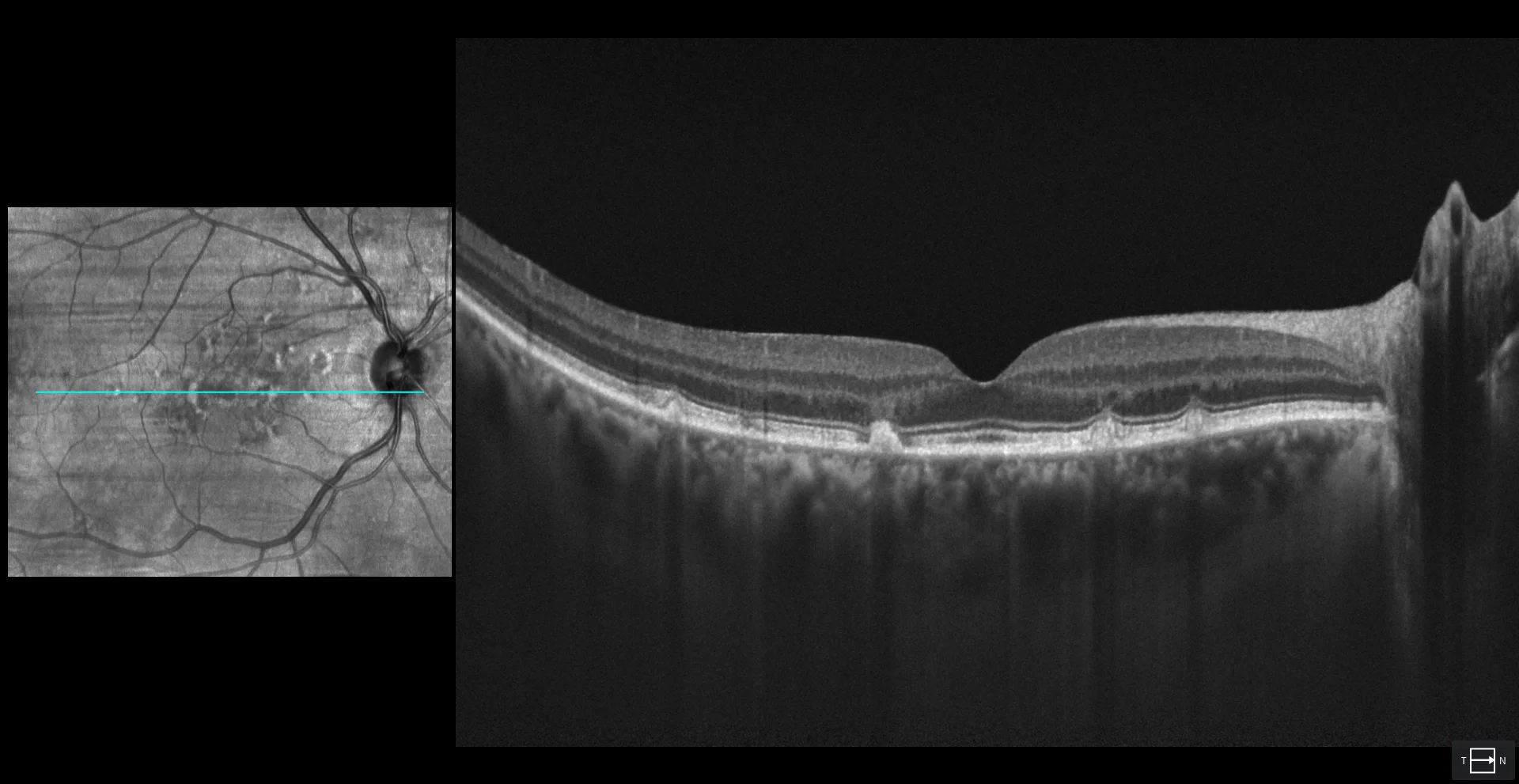
OCT (Cirrus 5000-HD, Zeiss): hyperreflective lesions above the RPE. Some are associated with disruption of the EZ and even the MLE, while in others these are intact. Some areas of thinning and atrophy of the RPE are also seen (C1, C2).
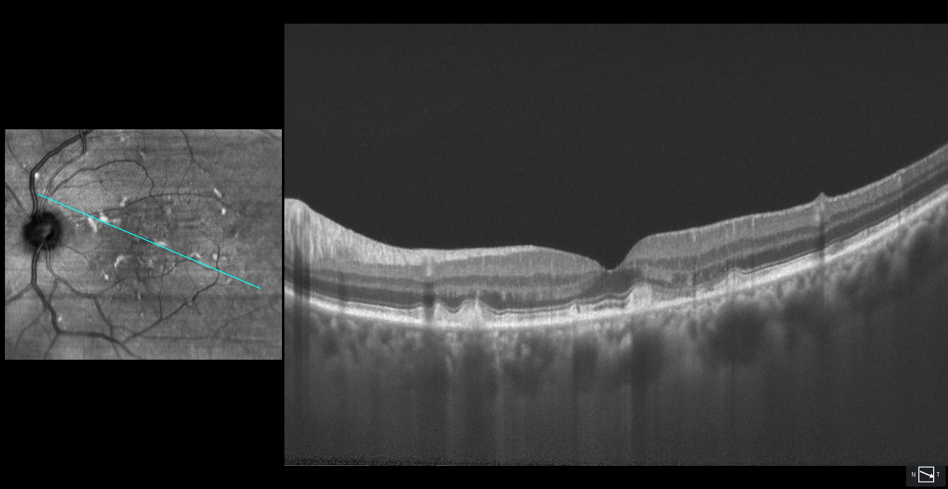
OCT (Cirrus 5000-HD, Zeiss): hyperreflective lesions above the RPE. Some are associated with disruption of the EZ and even the MLE, while in others these are intact. Some areas of thinning and atrophy of the RPE are also seen (C1, C2).
Description
A 65-year-old male comes in for a check-up.
VA OD 20/20 OS 20/20.
In the fundus examination, multiple small yellowish lesions are observed scattered throughout the macular area and around the optic disc. These lesions are hyperautofluorescent, suggesting they are vitelliform material, although there are also hypoautofluorescent areas due to RPE damage. On OCT, the lesions correspond to hyperreflective subretinal material, with some showing disruption of the ellipsoid zone (EZ). The appearance of the lesions, the high symmetry between both eyes, and the good visual acuity allow for the diagnosis of pattern dystrophy with a multifocal pattern mimicking Stargardt disease.