Peripapillary Intrachoroidal Cavitation
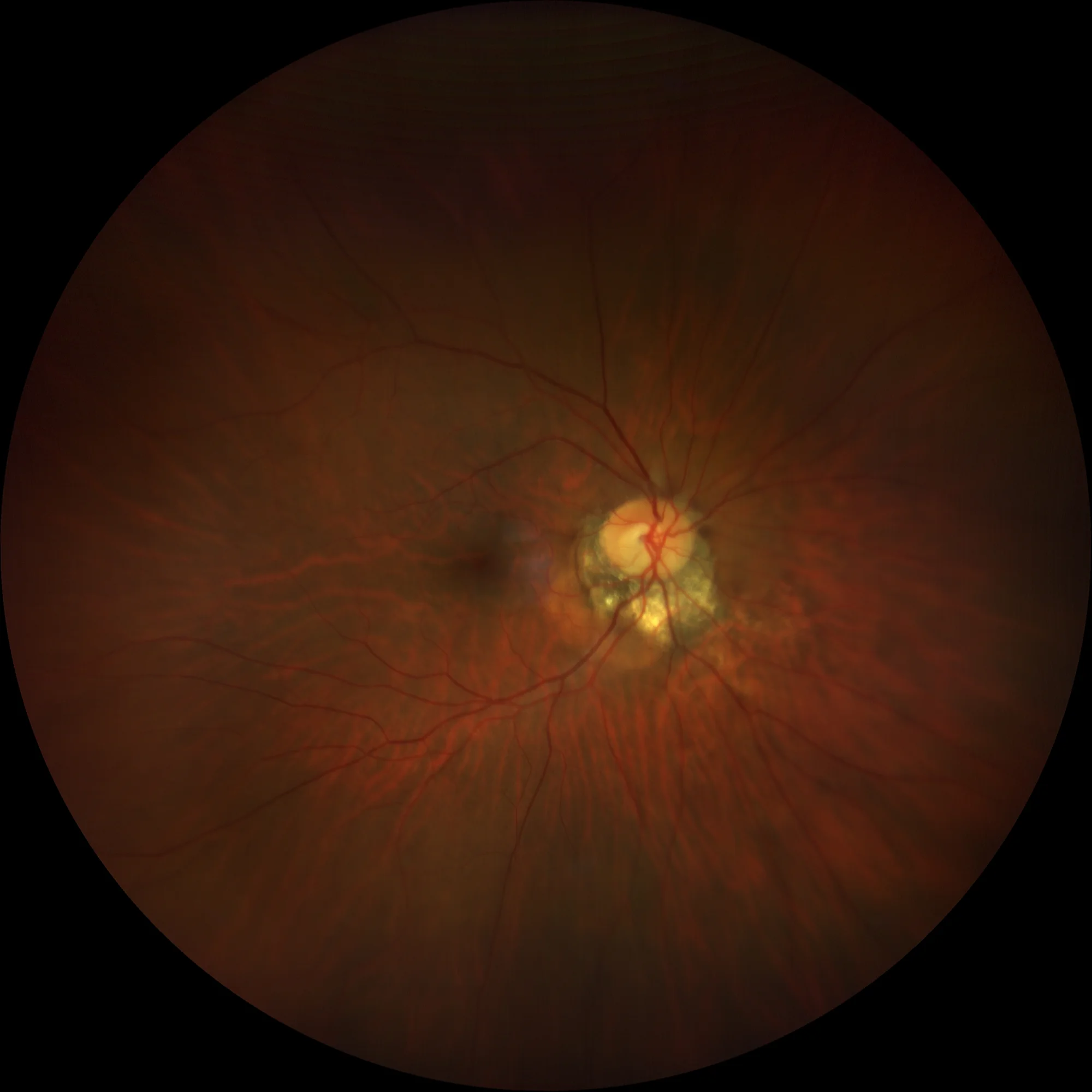
Intrachoroidal cavitation at the infero-temporal border of peripapillary atrophy.
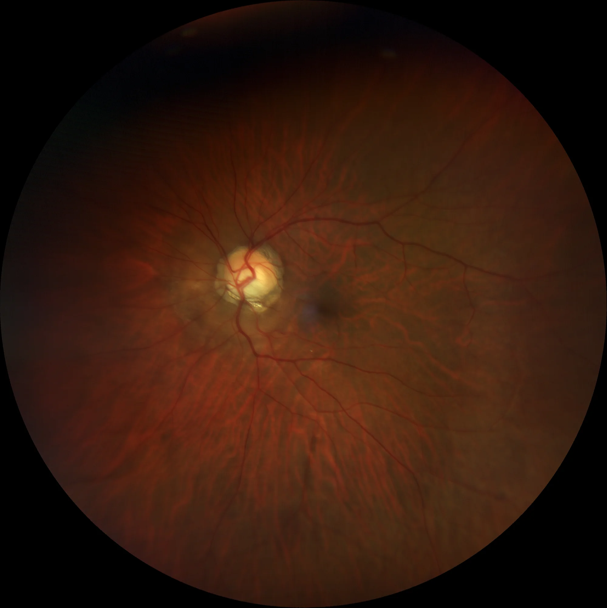
More extensive intrachoroidal cavitation than in the right eye, occupying approximately 2/3 of the papillary circumference.
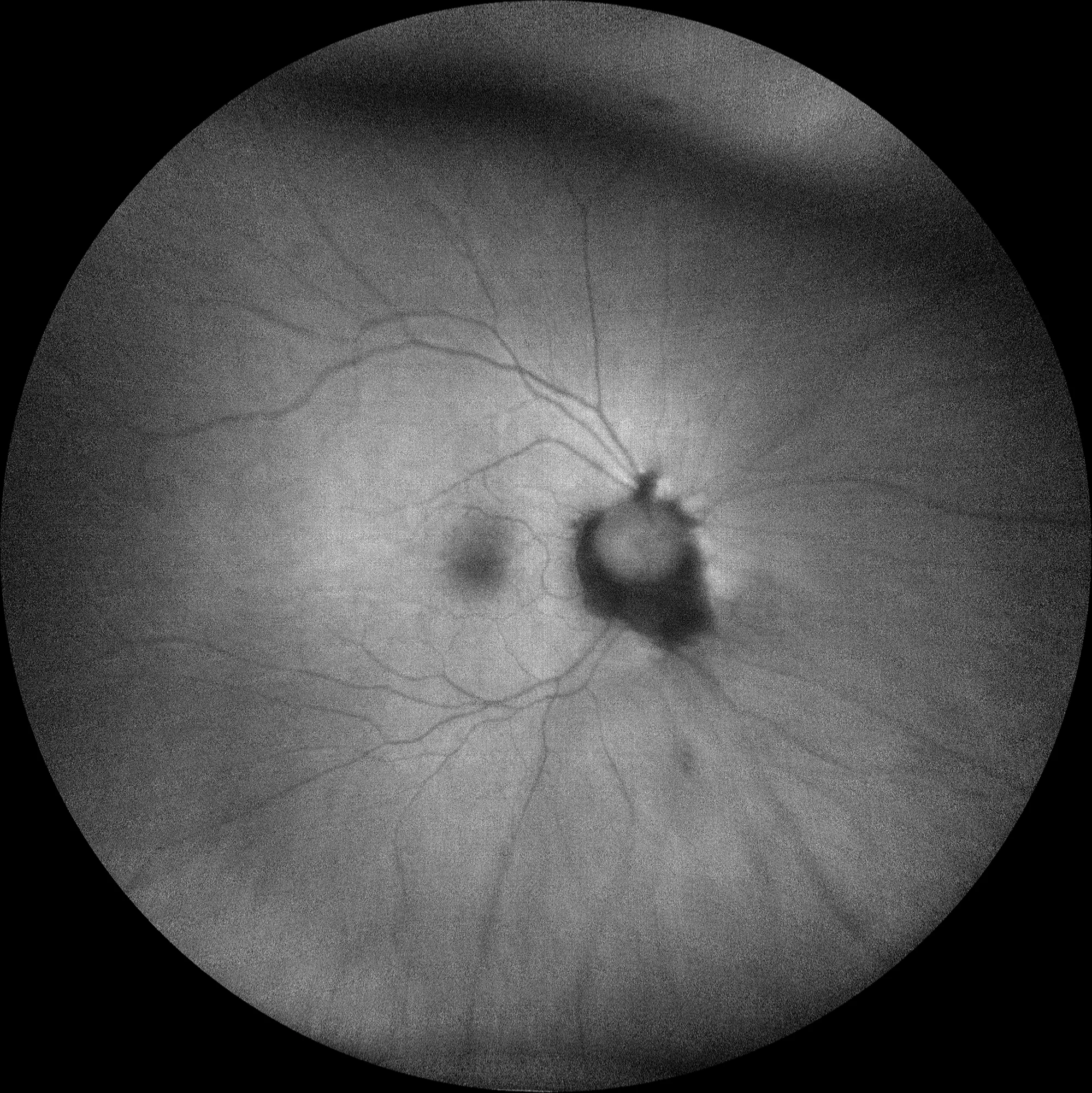
Autofluorescence (Clarus500, Zeiss):
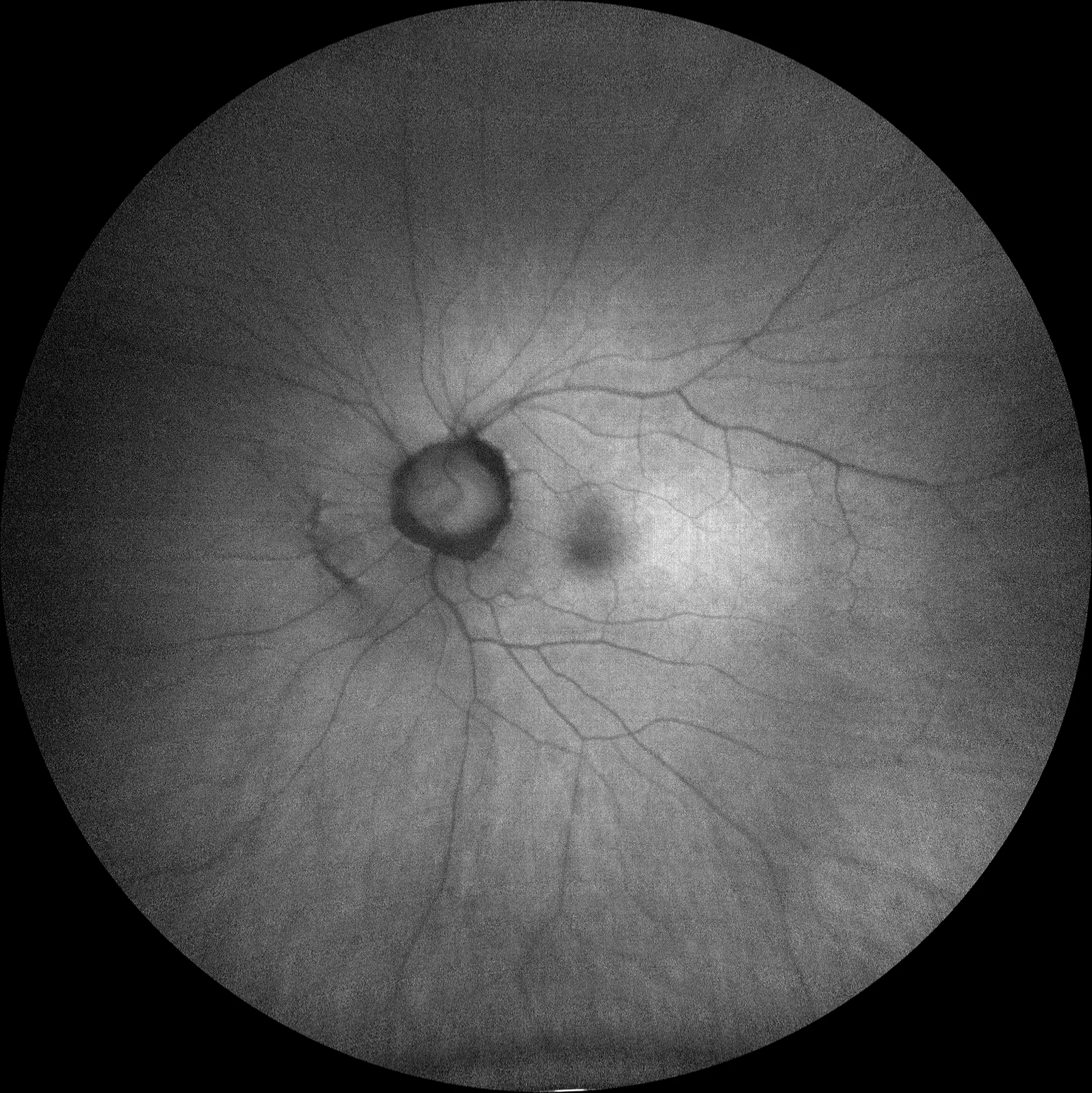
The autofluorescence shows peripapillary hypoautofluorescence corresponding to the peripapillary atrophy, without evident relevant alterations in the excavation area.
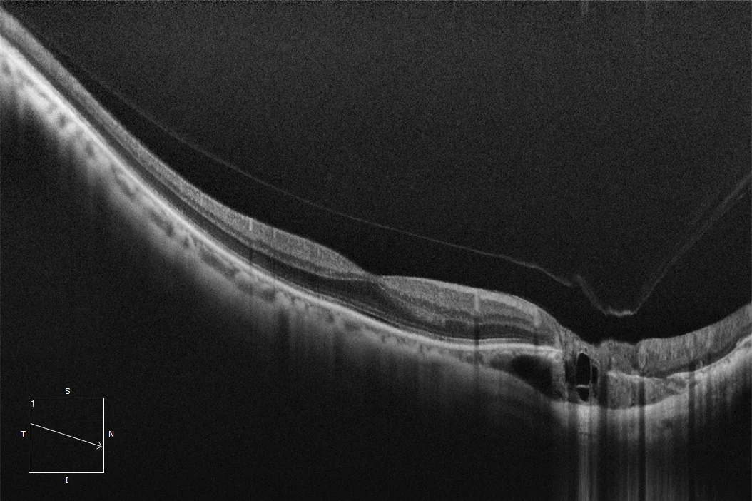
OCT of RE macula: Tilted macular plane with good foveal profile and choroidal thinning in the macular area. At the peripapillary level, choroidal thickening with cystic cavitations is observed, corresponding to the intrachoroidal cavitation.
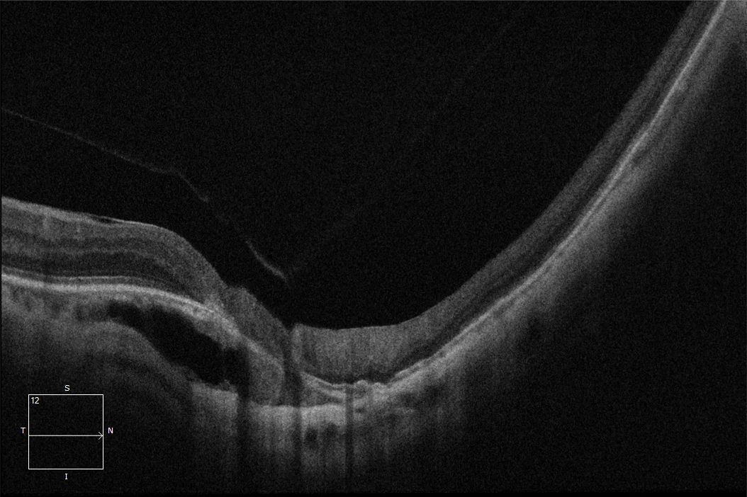
OCT over RE cavitation: The area of choroidal thickening with cystic cavities can be observed next to the peripapillary atrophy area.
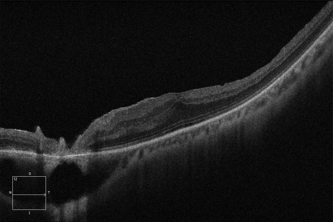
OCT of LE macula: Tilted macular plane. Thickened retina at the central level with loss of foveal profile due to history of ERM that required surgery. The cavitation in the peripapillary area can be observed.
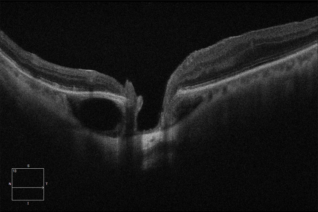
OCT over LE cavitation: Peripapillary choroidal thickening. In one of the sections, what appears to be a communication between the intrachoroidal cavitation and the vitreous cavity through the optic disc is observed.
Description
Well-defined yellow-orange lesion, located at the outer edge of the myopic cone (generally at the lower outer edge of the peripapillary gamma atrophy) and corresponding to an area of intrachoroidal thickening. These lesions may present intrachoroidal cysts and are associated with a discontinuity of the adjacent choroidal tissue. Its pathogenesis is unclear, but it is related to the posterior staphyloma. It has been considered a benign condition, but most patients present visual field defects and some may develop macular detachment with retinoschisis.