Peripapillary Intrachoroidal Cavitation
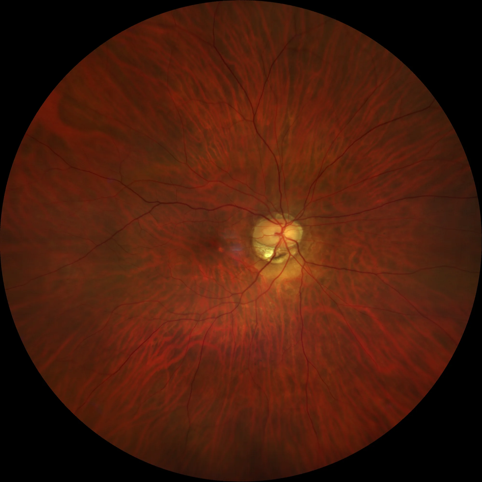
RE: Intrachoroidal cavitation at the lower edge of peripapillary atrophy. A small juxtafoveal atrophic lesion is observed.
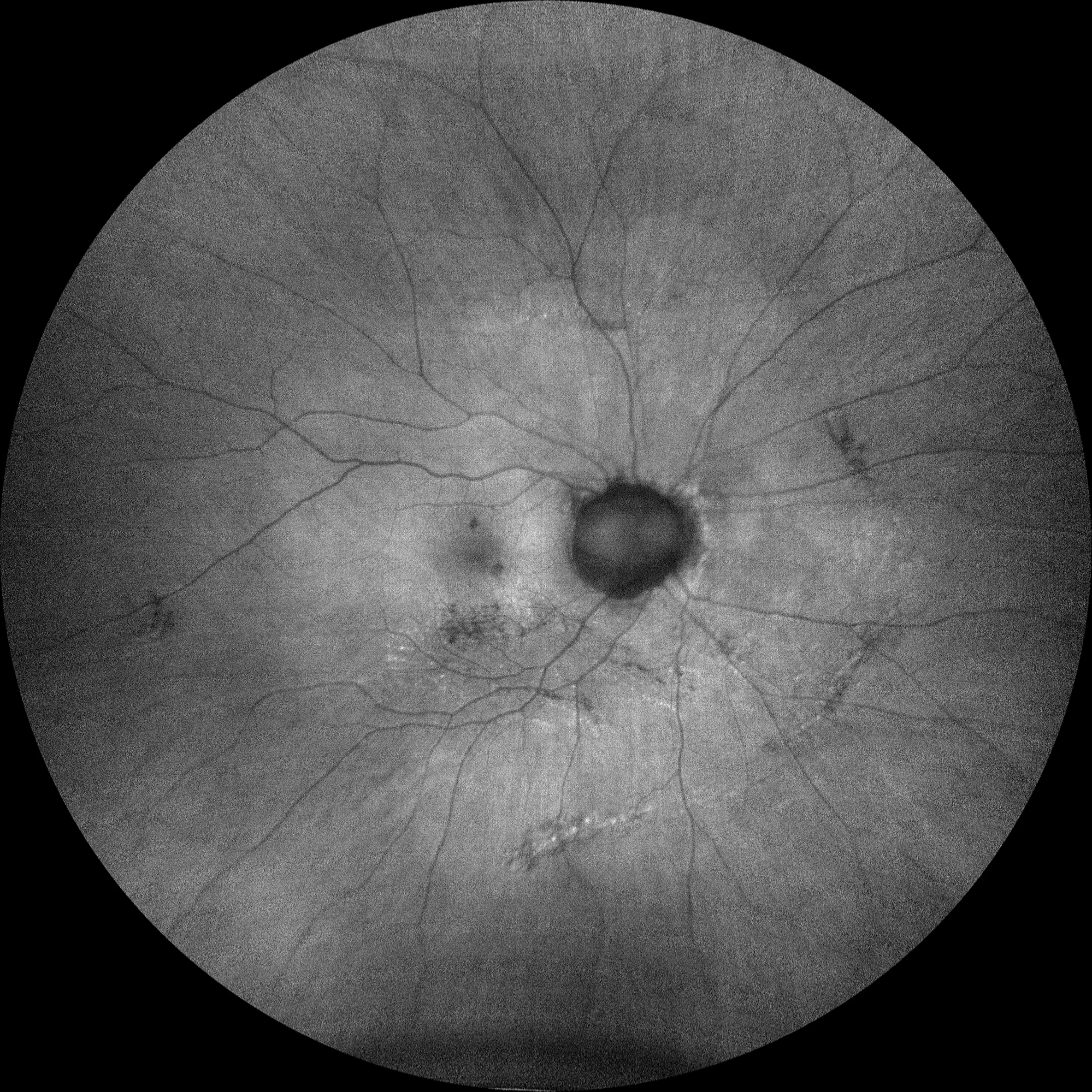
RE: Autofluorescence shows peripapillary hypoautofluorescence corresponding to peripapillary atrophy, with no significant alterations evident in the excavation area. A small juxtafoveal hypoautofluorescence is also observed in relation to the atrophic lesion visible in the retinography.
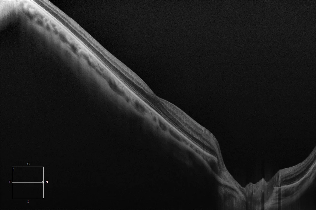
OCT macular RE: Tilted macular plane with good foveal profile and choroidal thinning in macular area. The cavitation is not observed as this is a cross-section passing superior to it, through the optic nerve and macula. (D) OCT over cavitation RE: The area of choroidal thickening with cystic cavities inferior to the optic disc can be observed.
Description
Well-defined yellow-orange lesion, located at the outer edge of the myopic cone (generally at the lower outer edge of the peripapillary gamma atrophy) and corresponding to an area of intrachoroidal thickening. These lesions may present intrachoroidal cysts and are associated with a discontinuity of the adjacent choroidal tissue. Its pathogenesis is unclear, but it is related to the posterior staphyloma. It has been considered a benign condition, but most patients present visual field defects and some may develop macular detachment with retinoschisis.