Proliferative diabetic retinopathy
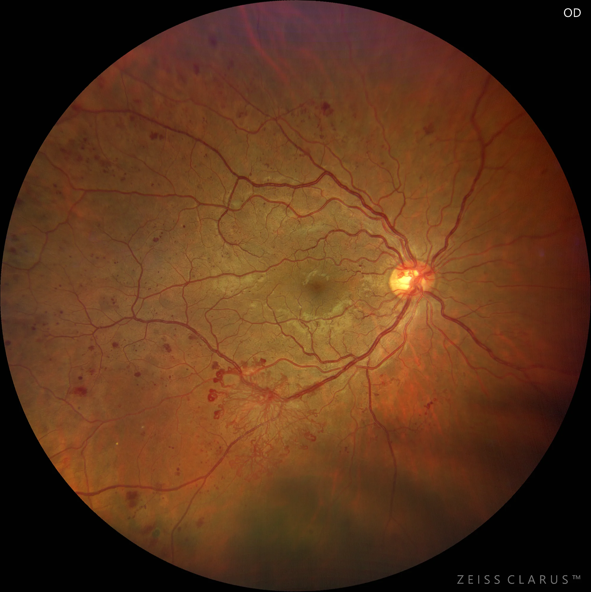
Color and red free WF retinography. Proliferative diabetic retinopathy with microaneurysms, retinal hemorrhages and neovascularization in the papilla and inferior temporal arch. The use of the green filter highlights the vascular alterations of PDR.
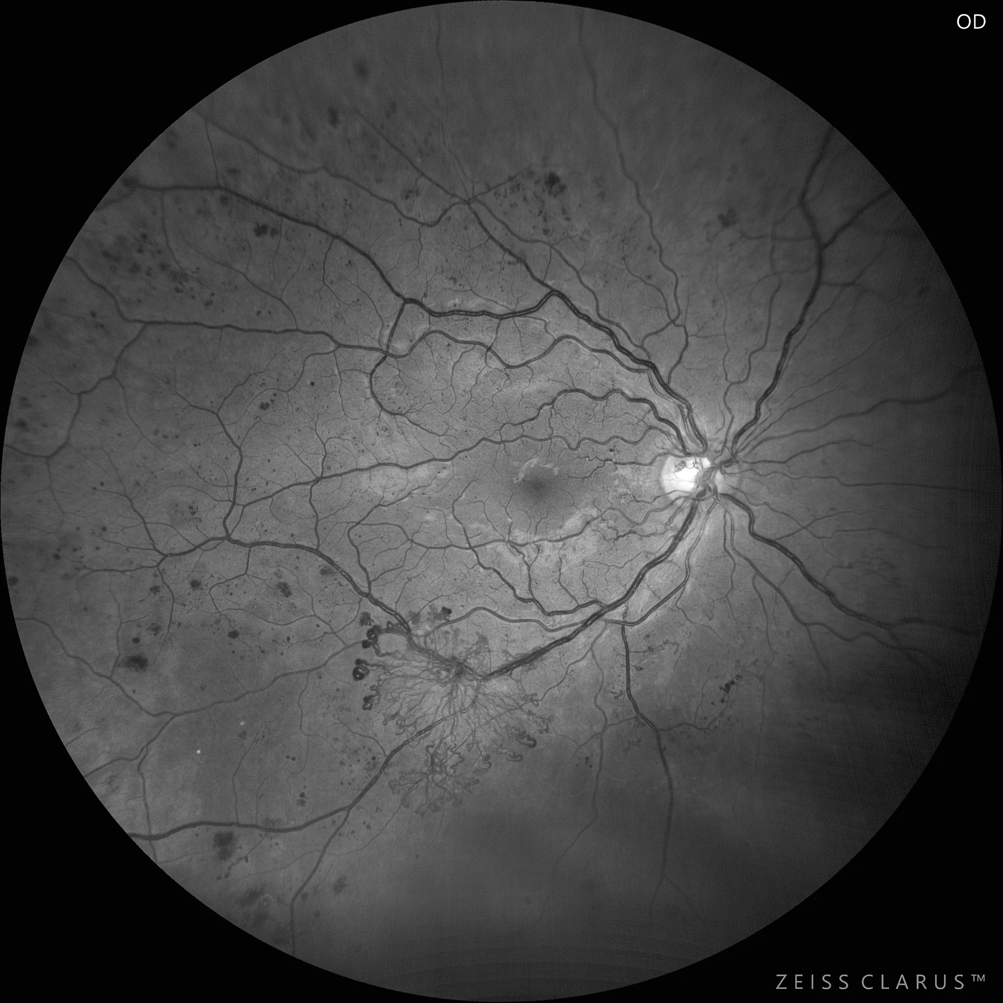
Color and red free WF retinography. Proliferative diabetic retinopathy with microaneurysms, retinal hemorrhages and neovascularization in the papilla and inferior temporal arch. The use of the green filter highlights the vascular alterations of PDR.
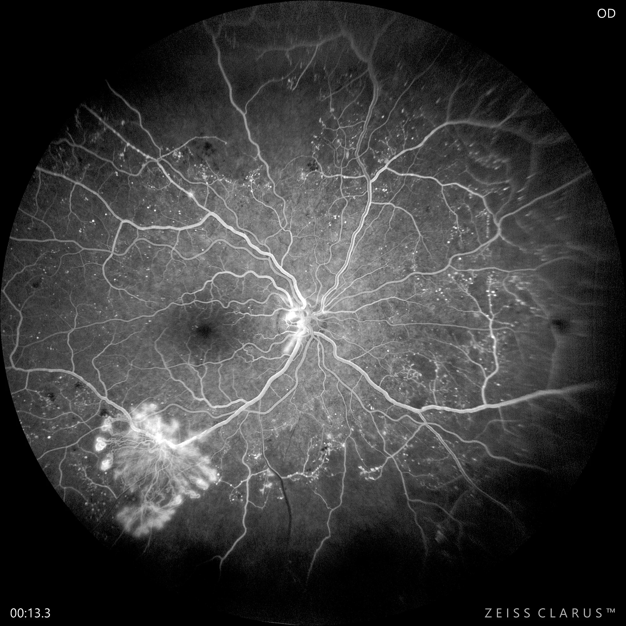
AGF WF. Confirms the presence of neovascularization in the papilla and inferior temporal arcade with marked dye oozing. Significant peripheral ischemia (initial and late phases).
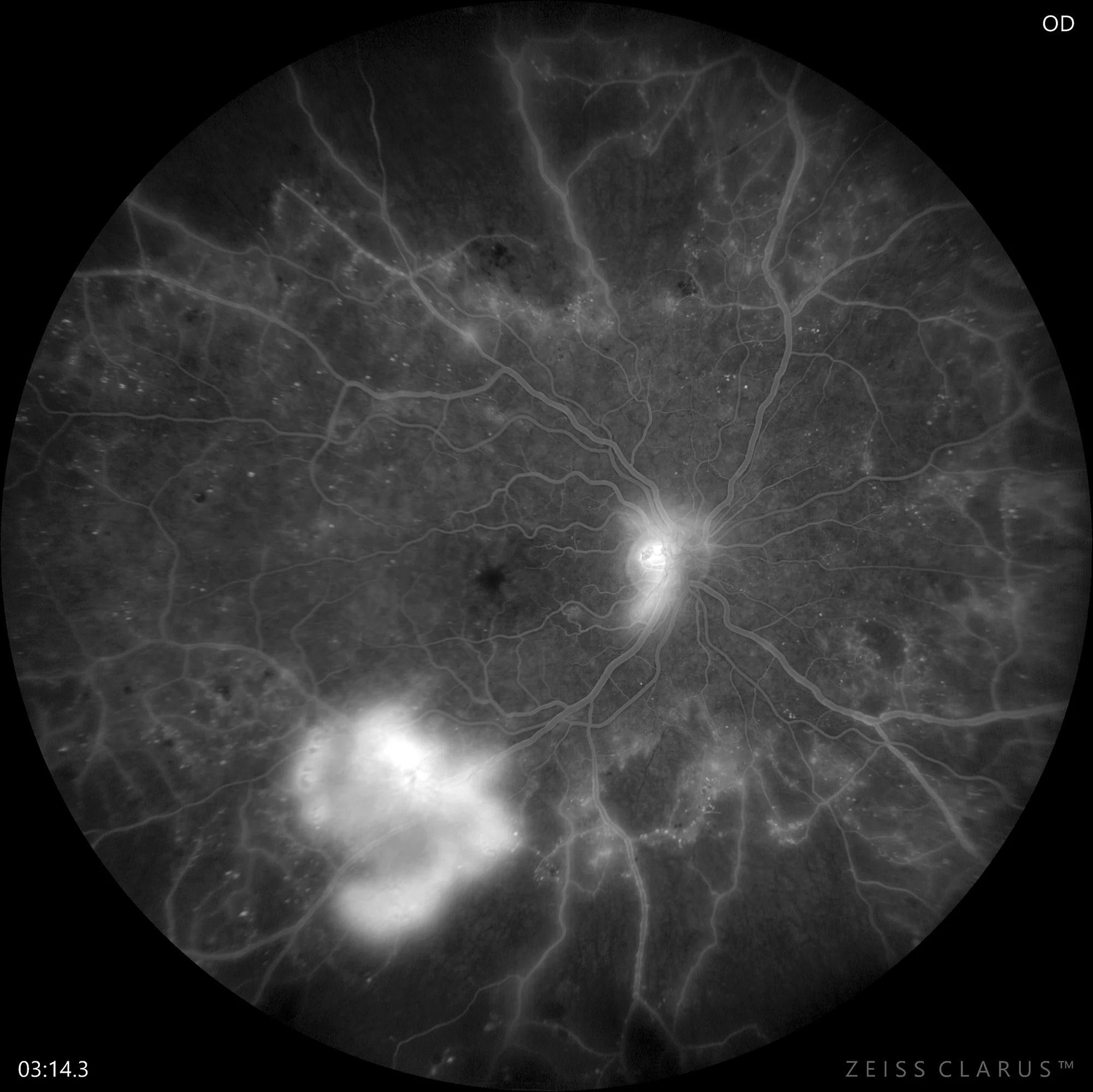
AGF WF. Confirms the presence of neovascularization in the papilla and inferior temporal arcade with marked dye oozing. Significant peripheral ischemia (initial and late phases).
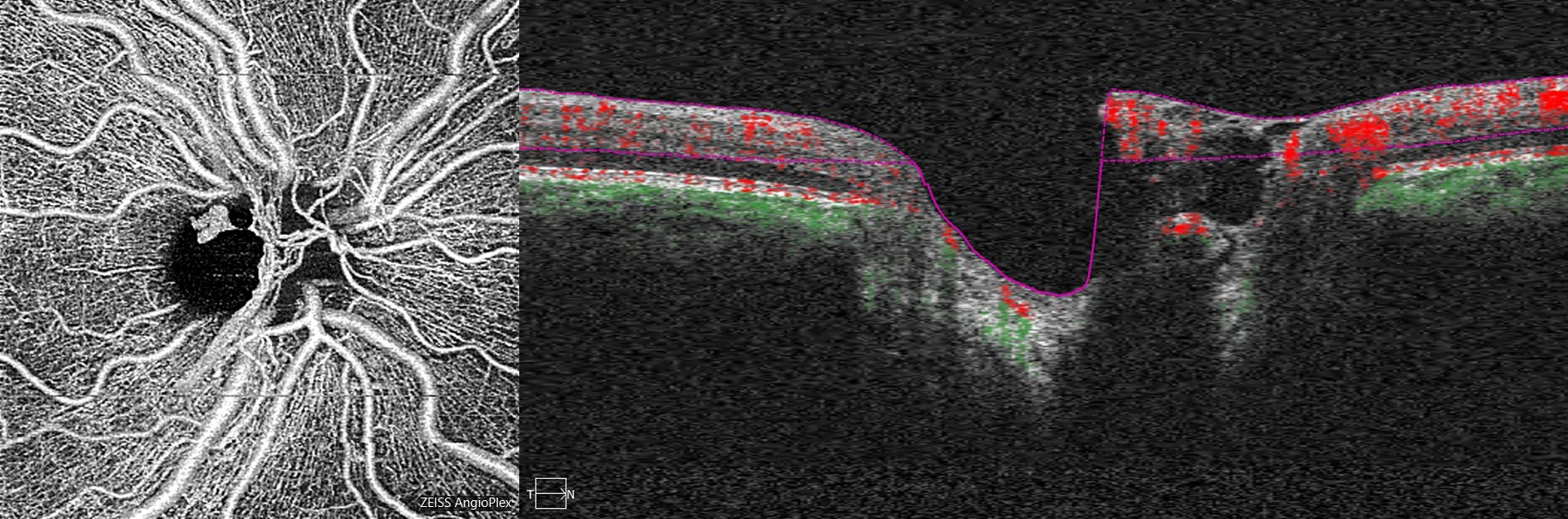
Angioplex (Cirrus 5000, Zeiss): Allows visualization of a detail of the papillary neovessels.
Description
Diabetic retinopathy is the most common retinal vascular disease. Proliferative diabetic retinopathy (PDR) is characterized by the appearance of neovascularization, constituting the most advanced phase of the disease. In these phases, vitreous hemorrhage is one of the main complications of sudden visual loss.