Racemose hemangioma
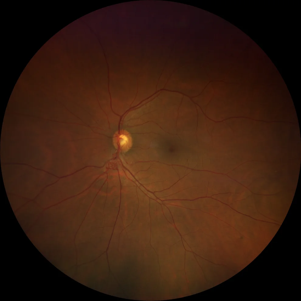
Color fundus (Clarus 500, Carl Zeiss Meditec ASG, Jena, Germany) of the left eye, showing an arteriovenous malformation around the exit of the inferior temporal vascular arcade.
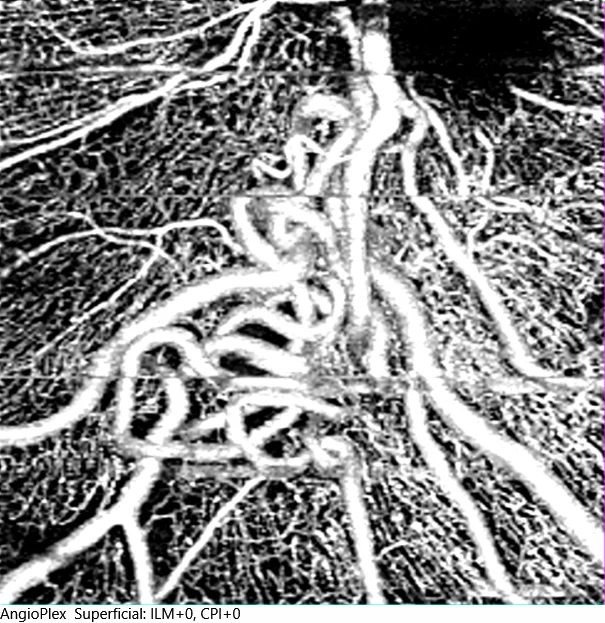
Optical coherence tomography angiography at the level of the superficial plexus (Cirrus 5000, Carl Zeiss Meditec ASG, Jena, Germany) of the arteriovenous malformation.
Description
Racemose hemangioma, also known as Wyburn-Mason syndrome, is a non-inherited congenital disease characterized by the presence of arteriovenous malformations (AVMs) in the retina, orbit, and brain. These malformations occur as abnormal connections between arteries and veins without the presence of an intermediate capillary network, resulting in direct, high-pressure blood flow.
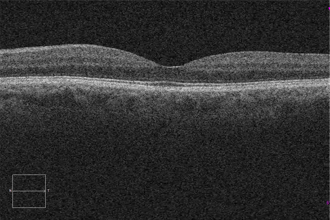
At the ocular level, retinal malformations (MAVs) are the main characteristic. These are usually unilateral and can affect both the retina and the optic nerve, typically presenting as dilated and tortuous vessels. Diagnosis is based on clinical identification and confirmation by imaging studies. AVMs can be detected by fluorescein angiography, revealing the abnormal flow of blood between arteries and veins. In addition, optical coherence tomography (OCT) can show thickening and secondary retinal edema.