Retinal Angiomatous Proliferation Stage 1
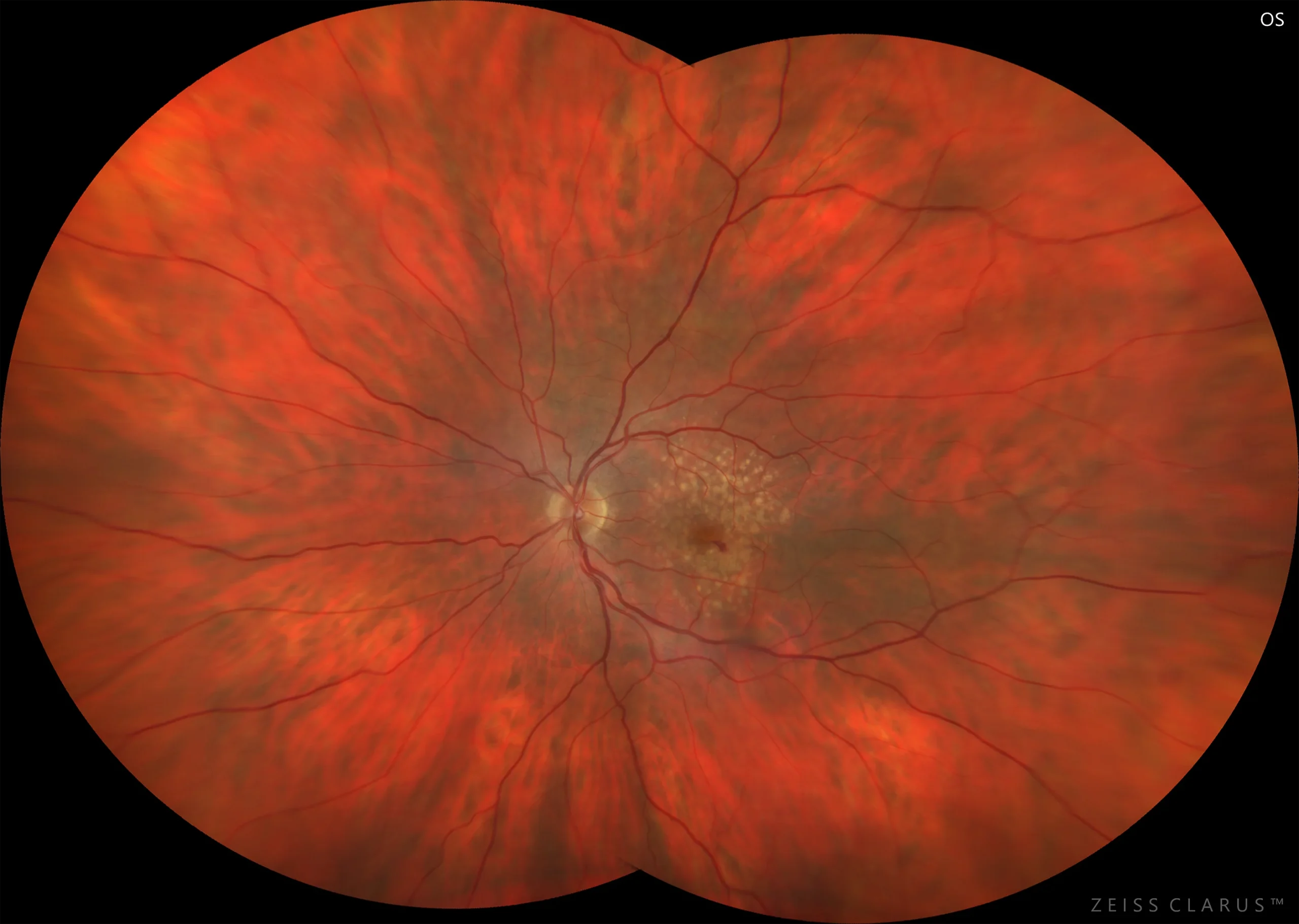
Figure 1. Color retinography of the left eye showing the presence of macular soft drusen and a small inferior juxtafoveal intraretinal hemorrhage.
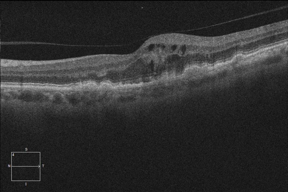
Figure 2. Optical Coherence Tomography. Multiple detachments of the drusenoid pigment epithelium. Intraretinal fluid.
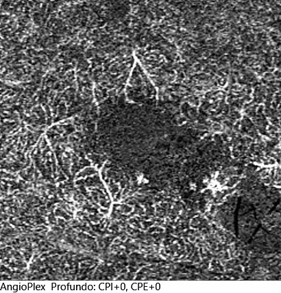
Figure 3. Angio-OCT of the deep plexus. Neovascular bouquet.
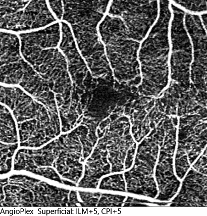
Figure 4. Angio-OCT of the superficial plexus. Increased inferior temporal vascular density
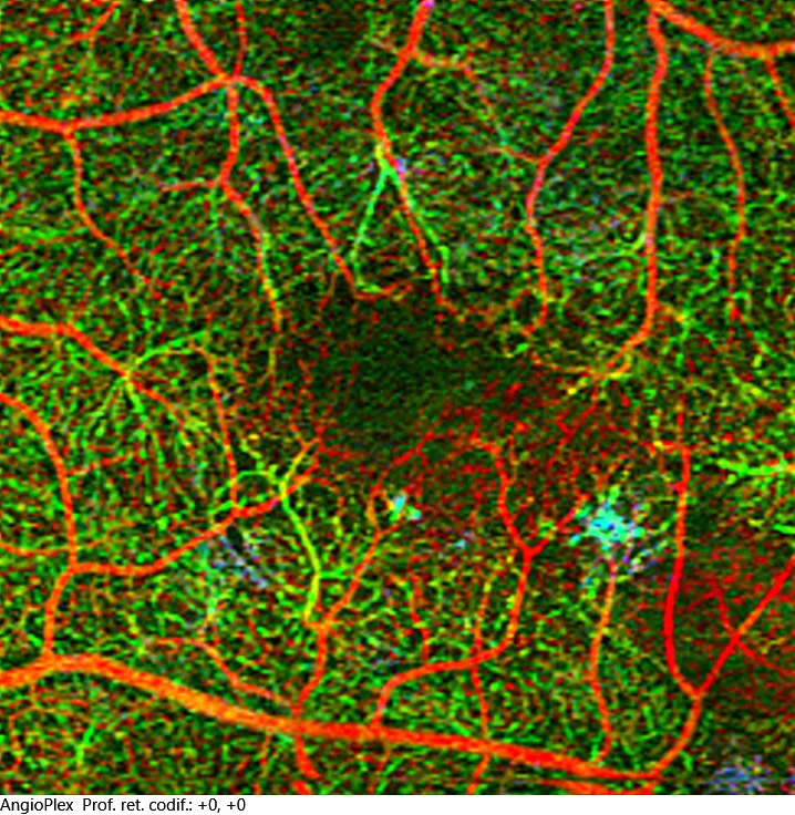
Figure 5. Face section of the deep plexus. Neovascular bunch
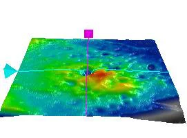
Figure 6. OCT with thickness map
Description
Characteristics:
The current classification of neovascular Age-Related Macular Degeneration (AMD) is based on the findings of Optical Coherence Tomography (OCT), which distinguishes 3 types of neovascular membrane, taking as an anatomical reference the line of the Retinal Pigment Epithelium (RPE) and the nature of the neovascular tissue.
Retinal angiomatous proliferation (RAP or AMD type 3) originates in the deep retinal vascular complex and is characterized by the presence of cystoid macular edema, which may develop into serous RPE detachment (PED), associated or not with subretinal fluid. The initial response to antiangiogenic treatment is very good, however, the high risk of development and progression to atrophy means that the long-term prognosis is not as good. Likewise, the high risk of bilaterality makes close monitoring of the contralateral eye necessary in order to make an early diagnosis.