Retinal dystrophy associated with mutation in the HK1 gene
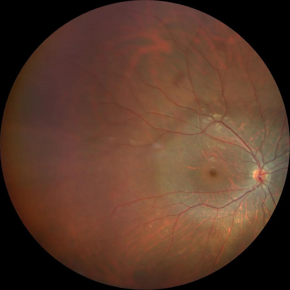
A and B. Color fundusography (Clarus 500, Carl Zeiss Meditec ASG, Jena, Germany) of the right and left eyes, showing retinal thinning and pallor with a circumferential distribution around the macula, following the vascular arcades.

A and B. Color fundusography (Clarus 500, Carl Zeiss Meditec ASG, Jena, Germany) of the right and left eyes, showing retinal thinning and pallor with a circumferential distribution around the macula, following the vascular arcades.
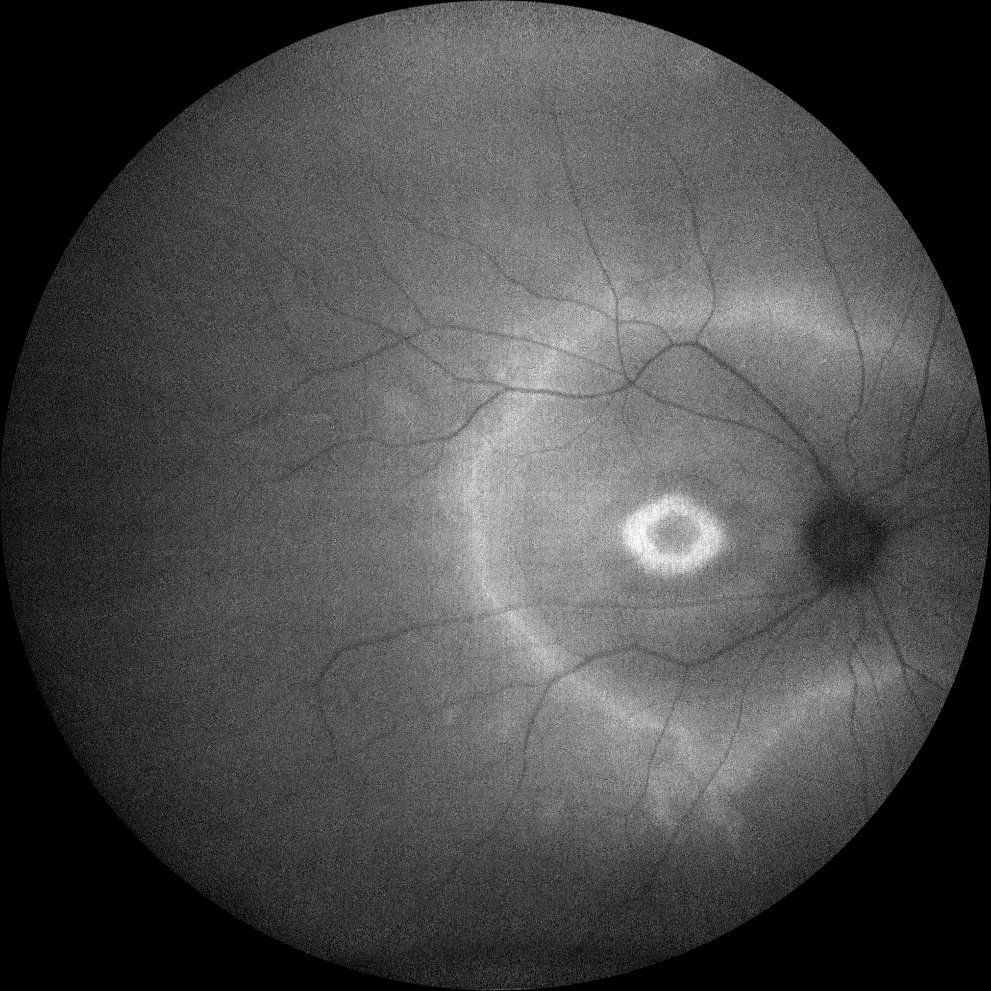
C and D. Autofluorescence images (Clarus 500, Carl Zeiss Meditec ASG, Jena, Germany) of the right and left eyes, showing a double hyperautofluorescent ring.
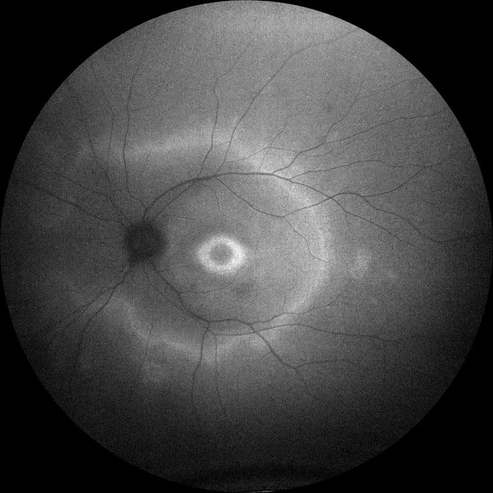
C and D. Autofluorescence images (Clarus 500, Carl Zeiss Meditec ASG, Jena, Germany) of the right and left eyes, showing a double hyperautofluorescent ring.
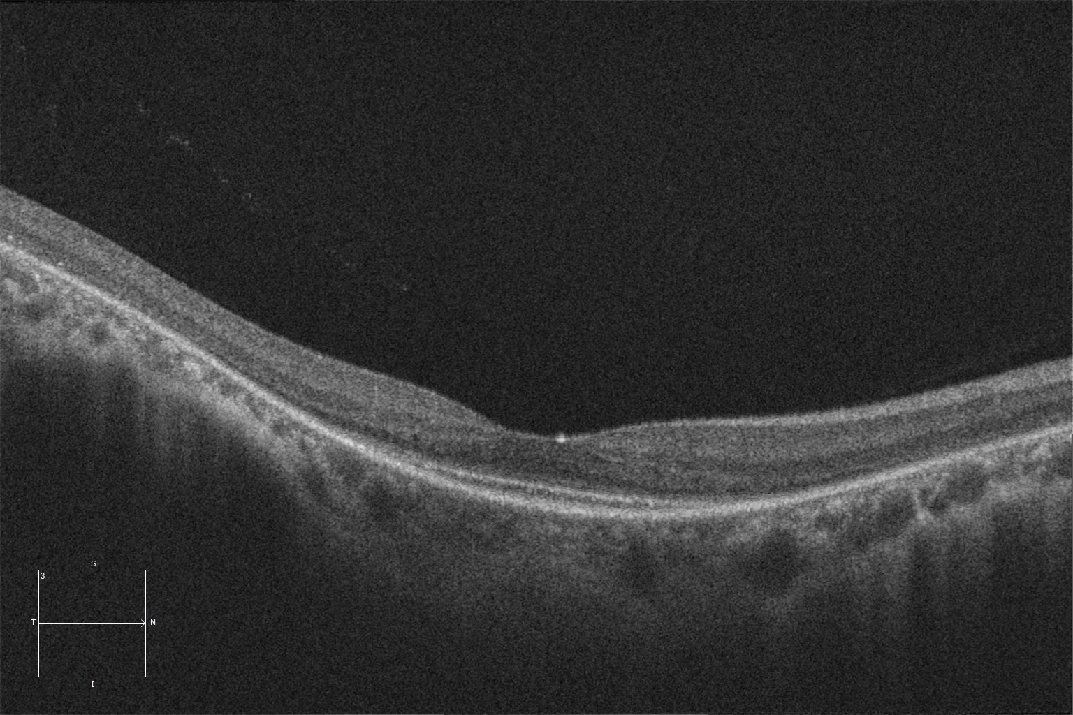
E and F. Macular HD optical coherence tomography (Cirrus 5000, Carl Zeiss Meditec ASG, Jena, Germany) of the right and left eyes, showing preservation of the ellipsoid and external limiting layers at the subfoveal level (within the internal hyperautofluorescent ring of autofluorescence), and absence of these retinal layers in the rest of the macula.
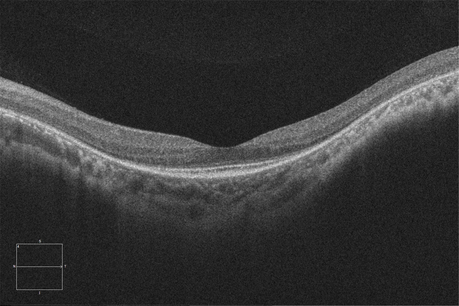
E and F. Macular HD optical coherence tomography (Cirrus 5000, Carl Zeiss Meditec ASG, Jena, Germany) of the right and left eyes, showing preservation of the ellipsoid and external limiting layers at the subfoveal level (within the internal hyperautofluorescent ring of autofluorescence), and absence of these retinal layers in the rest of the macula.
Description
Retinitis pigmentosa is a diffuse and hereditary retinal dystrophy that affects photoreceptors, initially rods and later cones (rod-cone dystrophy). The HK1 gene encodes for the prote in hexokinase 1, an important enzyme in the glycolysis pathway. Mutations in this gene have been associated with an autosomal dominant retinal dystrophy. Characteristically, the involvement in this dystrophy presents a double hyperautofluorescent ring, with preservation of photoreceptor layers inside the inner ring (foveal area) and outside the outer ring (peripheral retina), and absence of these layers in the area between the two rings. The penetrance in these families may be incomplete (i.e., patients carrying the pathogenic mutation may not present clinical signs of the disease). Among the most sensitive complementary tests for its diagnosis is the full-field electroretinogram, which will show a decrease in scotopic rod responses and subsequently, as the disease progresses, a decrease in photopic responses.
Comments
Hexokinase 1 is a ubiquitous enzyme; some recessive mutations in the same gene have been associated with hemolytic anemia without spherocytosis.Retinal dystrophy due to HK1 mutation. A and B. Color retinography (Clarus 500, Carl Zeiss Meditec ASG, Jena, Germany) of the right and left eyes, showing retinal thinning and pallor with a circumferential distribution around the macula, following the vascular arcades. C and D. Autofluorescence images (Clarus 500, Carl Zeiss Meditec ASG, Jena, Germany) of the right and left eyes, showing a double hyperautofluorescent ring. E and F. HD macular optical coherence tomography (Cirrus 5000, Carl Zeiss Meditec ASG, Jena, Germany) of the right and left eyes, showing preservation of the ellipsoid layer and external limiting membrane at the subfoveal level (within the internal hyperautofluorescent ring of the autofluorescence), and absence of these retinal layers in the rest of the macula.
Indication
25-year-old woman with nyctalopia since childhood. Her mother has a sim ilar clinical presentation. The genetic study reveals a pathogenic “missense” mutation in the HK1 gene.