Retinal pigment epithelium tear
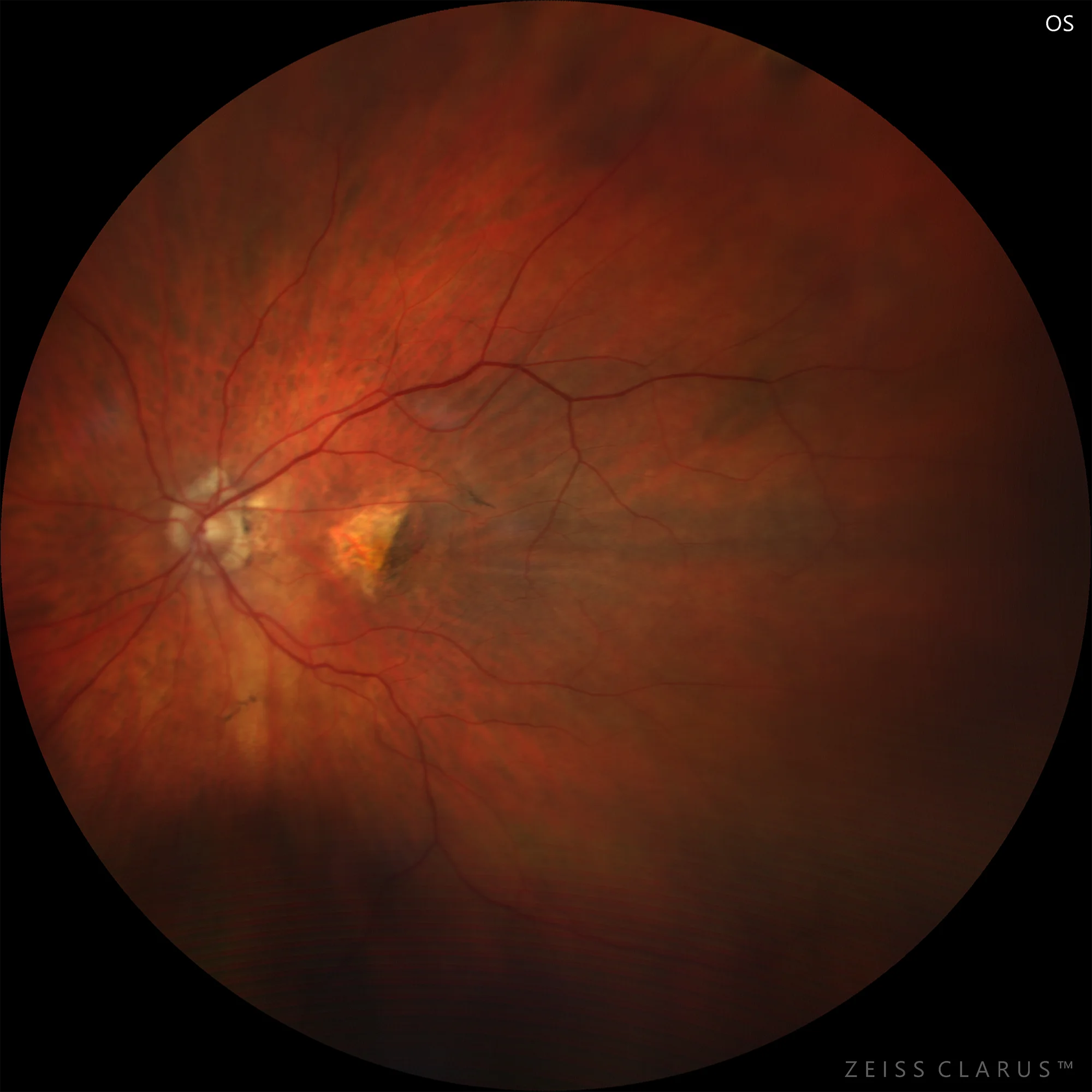
Figure 1. Color retinography showing the RPE tear and a fold. The choroid is shown denuded in the area of the tear.
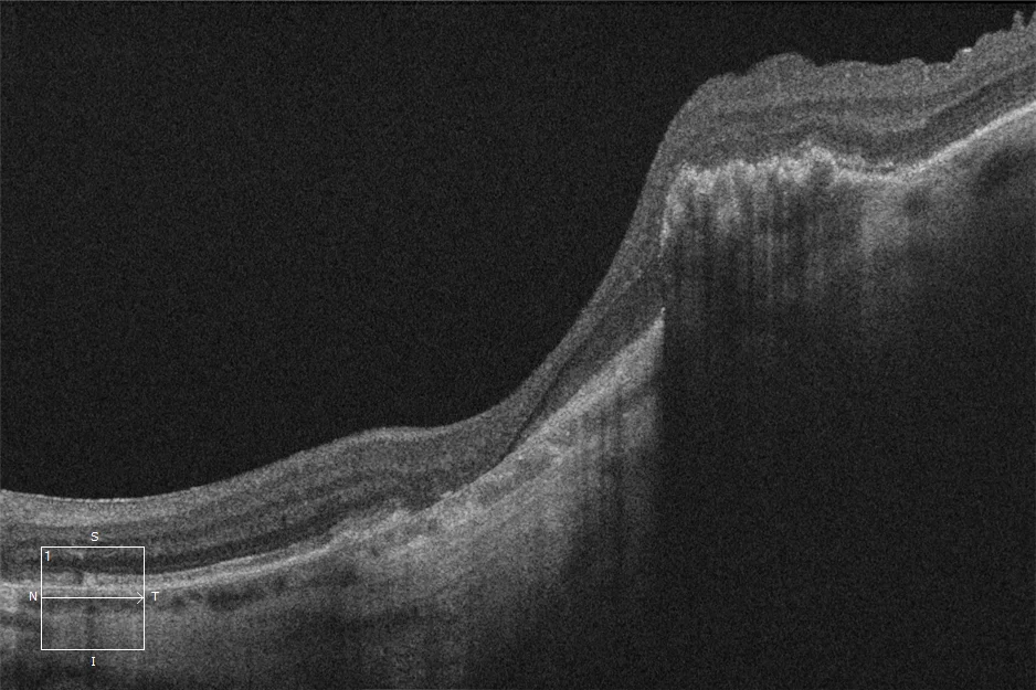
Figure 2. RPE band interruption. Hyperreflectivity in the area of the denuded choroid due to the greater penetration of the beam in the absence of the RPE shield.
Description
Retinal pigment epithelium (RPE) tear is a serious and rare complication of exudative age-related macular degeneration (AMD), which can occur spontaneously or after treatment with intravitreal therapy. It is usually associated with vascularized detachments of the RPE, and often involves a severe loss of visual acuity. The increase in the prevalence of AMD, as well as intravitreal treatments, are leading to a progressive increase in the incidence of this pathology.