Retinopathy of Prematurity
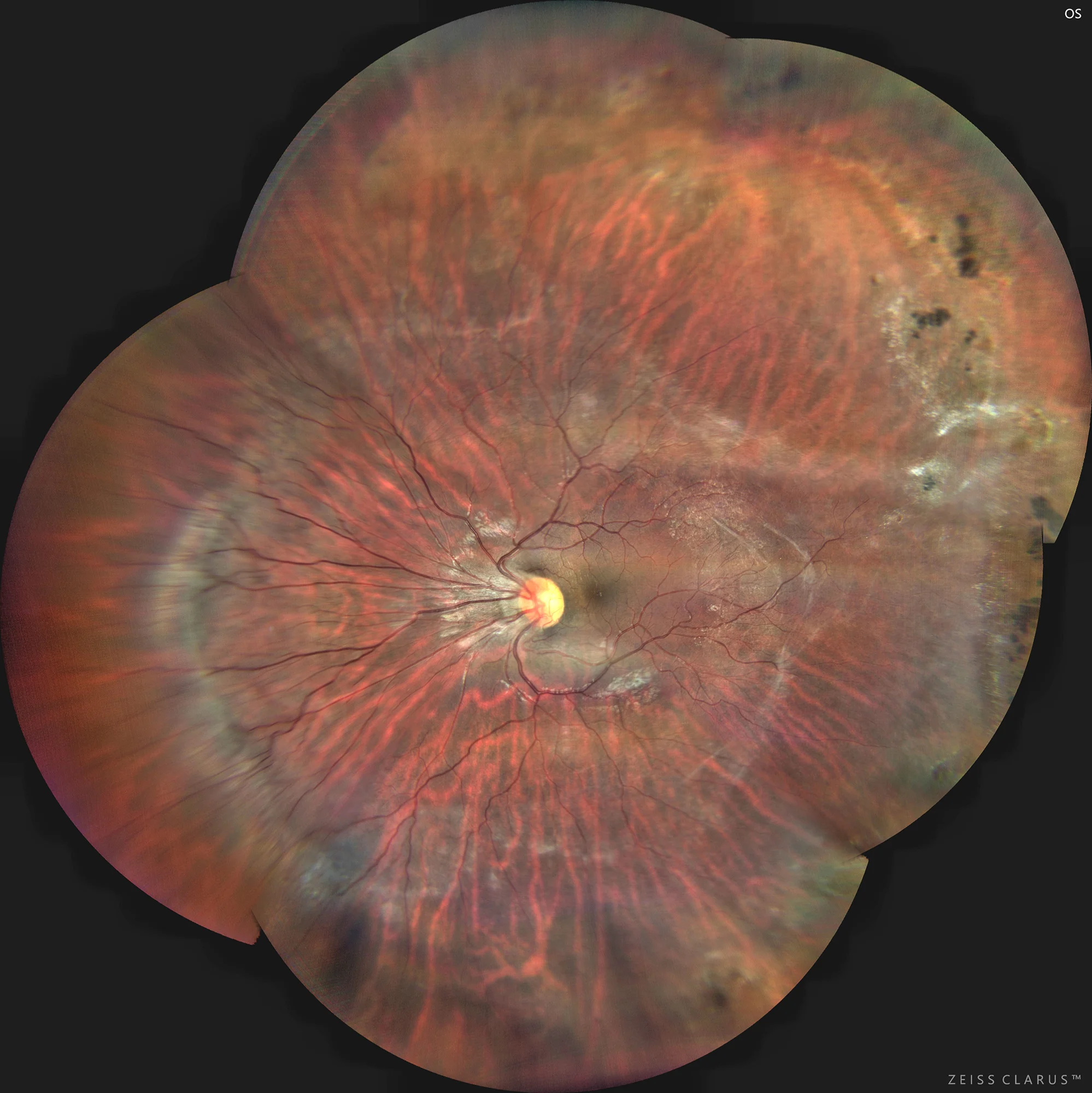
Color retinography showing multiple vascular abnormalities such as nasal deviation of vessels at the optic nerve exit, and elevation of vessels and the vitreoretinal interface on the nasal side forming a retinal ridge.
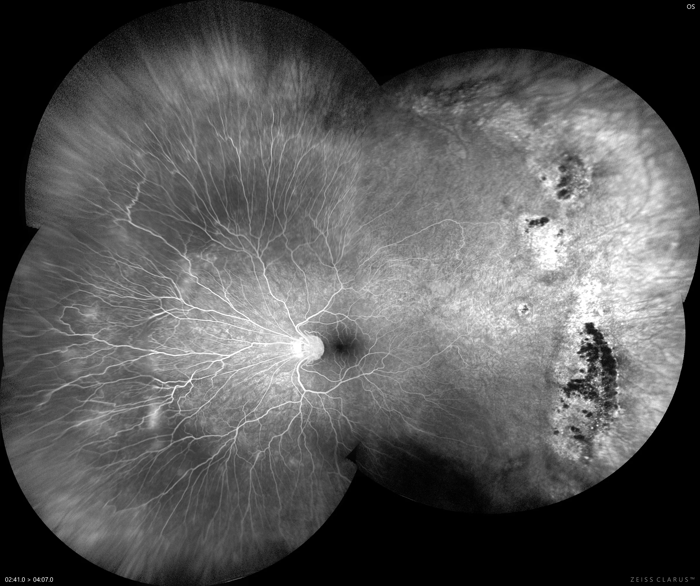
Fluorescein angiography: highlighting the presence of large avascular spaces in the temporal retina, along with hypofluorescent areas secondary to peripheral chorioretinal atrophy, where we previously observed strong vitreoretinal adhesion.
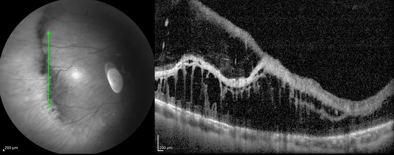
Peripheral OCT over the nasal ridge, in which we observe significant vitreoretinal traction, leading to schisis of the middle and inner retinal layers.
Description
Retinopathy of prematurity (ROP) in adulthood can present a variety of ocular complications due to the sequelae of the disease in its acute form. Adults with a history of ROP may develop significant myopia, strabismus, amblyopia, cataracts, glaucoma, and tractional retinal detachment. These conditions can arise due to vitreoretinal scarring and abnormalities in the vitreoretinal interface that result from the cicatricial phase of ROP. Additionally, it has been observed that patients with regressive ROP have a higher risk of presenting anomalies in foveal anatomy and macular pigmentation. Long-term monitoring is crucial to detect and treat these complications, as they can significantly affect vision.